
Review Article · Aesthetic Intelligence · Vol 1 · Issue 7
The Three-Cell Model of Skin RejuvenationThree Cells, One Framework
The fibroblast builds, the melanocyte must be respected, and the keratinocyte faces the world. Learn the three cells and every new booster, device or acid can be placed correctly the first time you meet it.
Dr Ahmed Haq1
- 1 Founder and Medical Director, Harley Street Institute, London, United Kingdom
Corresponding author: journal@harleystreetinstitute.com
Journal: Aesthet Intell
DOI: to be assigned
Volume / Issue: 1 / 7
Pages: 99–114
Received: 2026-06-18
Accepted: 2026-07-05
Published: 2026-07-07
Licence: CC BY 4.0
Abstract
- Background.
- Aesthetic skin practice is crowded with products, devices and protocols, yet the practitioner is rarely offered a simple organising principle that explains why any of them work. This review proposes a deliberately reductive teaching framework — the Three-Cell Model — which holds that almost every meaningful decision in non-surgical skin rejuvenation can be understood through the behaviour of just three resident cell populations: the fibroblast, the melanocyte and the keratinocyte.
- Methods.
- Narrative review and educational synthesis of established cutaneous cell biology and the clinical literature on retinoids, microneedling, energy-based devices, injectable biostimulators, chemical peels, photoprotection and topical actives, organised by cellular target rather than by product category.
- Results.
- The fibroblast is presented as the architect of the dermis, responsible for collagen, elastin and hyaluronic acid; the melanocyte as the pigment cell that governs evenness of tone and is the most reactive and least forgiving of the three; and the keratinocyte as the surface cell whose orderly maturation determines barrier function, hydration and light reflection. For each cell, the model maps the common clinical interventions onto the specific cellular behaviour they modulate. The model also carries a safety corollary: because the melanocyte responds to insult with pigmentary chaos, any intervention that risks provoking it must be approached with particular caution in higher Fitzpatrick skin types.
- Conclusion.
- The purpose of the model is not to replace detailed cell biology but to give the practitioner, and above all the trainee, a durable mental scaffold onto which new treatments can be placed as they emerge. The Three-Cell Model is offered as an educational tool, not a biological claim of completeness.
Keywords: skin rejuvenation, fibroblast, melanocyte, keratinocyte, retinoids, aesthetic education, collagen, skin barrier
| Evidence domain | What the article reports | How to interpret it |
|---|---|---|
| Background | Aesthetic skin practice is crowded with products, devices and protocols, yet the practitioner is rarely offered a simple organising principle that explains why any of them work. This review proposes a deliberately reductive teaching framework — the Three-Cell Model — which holds that almost every meaningful decision in non-surgical skin rejuvenation can be understood through the behaviour of just three resident cell populations: the fibroblast, the melanocyte and the keratinocyte. | Defines the problem and the reason for review or investigation. |
| Methods | Narrative review and educational synthesis of established cutaneous cell biology and the clinical literature on retinoids, microneedling, energy-based devices, injectable biostimulators, chemical peels, photoprotection and topical actives, organised by cellular target rather than by product category. | Shows how the evidence or observations were assembled. |
| Results | The fibroblast is presented as the architect of the dermis, responsible for collagen, elastin and hyaluronic acid; the melanocyte as the pigment cell that governs evenness of tone and is the most reactive and least forgiving of the three; and the keratinocyte as the surface cell whose orderly maturation determines barrier function, hydration and light reflection. For each cell, the model maps the common clinical interventions onto the specific cellular behaviour they modulate. The model also carries a safety corollary: because the melanocyte responds to insult with pigmentary chaos, any intervention that risks provoking it must be approached with particular caution in higher Fitzpatrick skin types. | Summarises the principal findings reported by the authors. |
| Conclusion | The purpose of the model is not to replace detailed cell biology but to give the practitioner, and above all the trainee, a durable mental scaffold onto which new treatments can be placed as they emerge. The Three-Cell Model is offered as an educational tool, not a biological claim of completeness. | States the authors’ clinical interpretation and its limits. |
Postgraduate Level
Complete this article to earn your certificate
Read the article, complete a short assessment, and submit your reflection to receive your AiCE Points certificate. Equivalence to 1 CPD/CME point — we do not award CPD/CME directly.
Learning Objectives
- 1Explain the roles of the fibroblast, melanocyte and keratinocyte in skin quality and ageing
- 2Map retinoids, microneedling, energy-based devices and biostimulators onto fibroblast stimulation
- 3Calibrate inflammatory treatments to Fitzpatrick phototype to avoid post-inflammatory hyperpigmentation
- 4Distinguish barrier support and turnover regulation as keratinocyte-directed strategies
- 5Apply the order of operations: dermal foundation, pigment protection, then surface refinement
The Three-Cell Model — Quick Reference
Cell 1 · Fibroblast (the architect)
- Location
- Dermis.
- Output
- Collagen, elastin, hyaluronic acid.
- Failure mode
- Goes quiet — synthesis falls, MMP-driven breakdown dominates.
- Aim
- Stimulate — retinoids, microneedling, energy devices, biostimulators, polynucleotides.
Cell 2 · Melanocyte (the temperamental one)
- Location
- Basal layer of the epidermis.
- Output
- Melanin — tone, evenness, endogenous UV protection.
- Failure mode
- Responds to insult with uneven pigment; melasma, PIH.
- Aim
- Calm and protect — tyrosinase inhibitors, photoprotection; de-escalate in Fitzpatrick IV–VI.
Cell 3 · Keratinocyte (the surface)
- Location
- Epidermis, maturing upward to the stratum corneum.
- Output
- Barrier function, hydration, light reflection (“glow”).
- Failure mode
- Disordered turnover — dullness, roughness, raised TEWL.
- Aim
- Regulate turnover and support the barrier — peels, retinoids, ceramide-based topicals.
Cross-cutting
- Acts on two cells
- Retinoid — fibroblast and keratinocyte.
- Protects all three
- Daily broad-spectrum photoprotection.
- Order of operations
- Dermal foundation → respect pigment → refine surface.
1. Introduction
A practitioner beginning a career in aesthetic skin medicine faces a peculiar problem. There is no shortage of information — there are thousands of products, dozens of device categories, and a continuous stream of new injectable “boosters,” each accompanied by its own marketing and its own supposedly unique mechanism. What is genuinely scarce is a simple framework that explains how any of it fits together. The result is that many practitioners learn skin treatments as a list of branded protocols to be memorised, rather than as a small number of biological principles to be understood.[1]
This review sets out a teaching model that I have used for many years in postgraduate aesthetic training. It rests on a single simplifying claim: that the overwhelming majority of what we do to rejuvenate skin can be understood through the behaviour of three cells. If a practitioner understands what the fibroblast, the melanocyte and the keratinocyte each do, why each one falters with age or injury, and which of our interventions acts on which cell, then almost any new treatment can be placed correctly the first time it is encountered.
The model is intentionally reductive. Human skin contains many more cell types than three — Langerhans cells, Merkel cells, mast cells, endothelial cells, adipocytes and the immune infiltrate all matter, and none is dismissed here.[2] The claim is not that three cells are all that exist, but that three cells are enough to organise the practitioner's thinking. In education, a framework that is remembered and used is worth more than a complete one that is not. What follows describes each cell in turn, in plain language, and then maps our common treatments onto them.
2. The architecture of the skin, in brief
Before the cells, a word on the ground they occupy, because the model depends on it. Skin is arranged in layers. The outermost is the epidermis, a thin, cell-dense sheet that faces the world. Beneath it lies the dermis, a thicker, tougher layer that provides mechanical strength and holds the structures — vessels, nerves, glands — on which the skin depends. Below the dermis sits the subcutaneous fat.[3]
The single most useful fact for the practitioner is that water and nutrients diffuse upward, from the vascularised dermis toward the surface. The epidermis has no blood supply of its own; it is sustained from below.[3,4] This simple directionality explains a great deal of what follows, and it is the reason this review treats the cells in a specific order — the deep, generative fibroblast first, then upward to the surface keratinocyte — rather than the conventional outside-in sequence of the histology textbook.
3. Cell one: the fibroblast, the architect
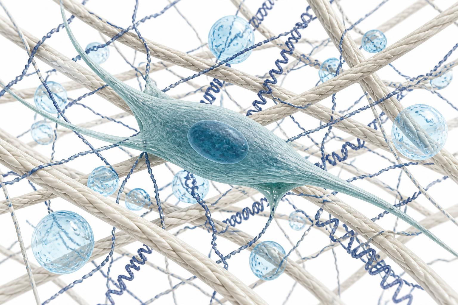
The fibroblast lives in the dermis, and it is the closest thing the skin has to a builder. It manufactures the three materials that give young skin its qualities: collagen, which provides tensile strength and structure; elastin, which provides recoil; and hyaluronic acid, a water-binding molecule that provides volume and hydration.[5,6] When people describe skin as “firm,” “bouncy” or “plump,” they are describing, in lay terms, the output of well-functioning fibroblasts.
The trouble is that fibroblast activity declines with age. From the third decade onward, collagen synthesis falls, and the balance between production and breakdown tips toward breakdown, driven in part by matrix metalloproteinases — enzymes that degrade the existing collagen scaffold.[7,8] Ultraviolet exposure accelerates this process dramatically; the majority of what patients experience as “ageing” of the skin is in fact photoageing, a cumulative, sun-driven degradation of the dermal matrix.[9,10] The fibroblast, in effect, goes quiet, and the architecture it once maintained is left to decay without adequate repair.
Almost everything we do to “stimulate,” “regenerate” or “rebuild” skin is an attempt to wake the fibroblast up. This is the unifying insight of the entire category. Retinoids — vitamin A derivatives such as tretinoin — are among the most evidence-supported topical agents in dermatology; they increase collagen production, normalise the skin's turnover and reduce the enzymes that break collagen down.[11,12] A practitioner who understands the fibroblast understands immediately why the retinoid is foundational rather than optional.
Microneedling creates controlled micro-injuries in the dermis; the wound-healing response that follows recruits fibroblasts and stimulates new collagen.[13] Energy-based devices — fractional lasers, radiofrequency, ultrasound — deliver thermal or ablative injury to the dermis, again provoking a fibroblast-mediated repair response and neocollagenesis.[14,15] Injectable biostimulators, such as poly-L-lactic acid and calcium hydroxylapatite, act not by filling space directly but by prompting the fibroblast to lay down new collagen around the injected material over subsequent weeks and months.[16,17] Polynucleotides, a more recent category, are proposed to support fibroblast activity and dermal hydration, with early randomised evidence of improvement in skin quality parameters.[18]
Six different interventions; one cellular target. The practitioner who has learned the fibroblast does not need to memorise six unrelated protocols. They need to ask a single question of any new treatment: does this wake the fibroblast, and if so, how forcefully?
4. Cell two: the melanocyte, the temperamental one
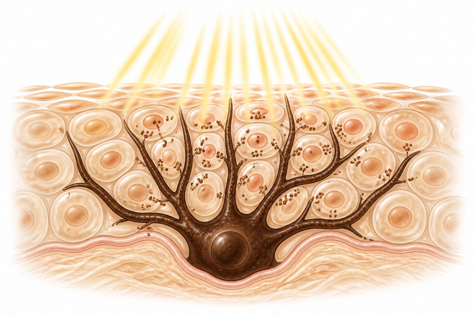
The melanocyte sits at the base of the epidermis, at the junction with the dermis, nestled among the basal stem cells.[19] Its job is to produce melanin, the pigment that gives skin its colour and, importantly, protects the deeper cells from ultraviolet damage by absorbing and scattering radiation. Melanin is, in evolutionary terms, the skin's own sunscreen.[20]
For the aesthetic practitioner, the melanocyte matters for a different reason: it is the cell responsible for evenness of tone, and it is the least forgiving cell in the skin. When a melanocyte is provoked — by ultraviolet light, by inflammation, by hormonal change, or by clumsy treatment — it does not fail quietly like the fibroblast. It responds by producing pigment, often unevenly, producing the patches, spots and diffuse discolouration that patients describe as “pigmentation.”[21] Conditions such as melasma and post-inflammatory hyperpigmentation are, at root, disorders of a melanocyte that has been pushed too hard.[22]
This gives the model its most important safety lesson. Any treatment that causes inflammation or injury — a peel, a laser, even an aggressive microneedling session — carries a risk of provoking the melanocyte into post-inflammatory hyperpigmentation. That risk is not evenly distributed: it rises substantially with higher Fitzpatrick skin phototypes (IV, V and VI), in which the melanocyte is more reactive.[23,24] The practical consequence is that the practitioner must calibrate the aggressiveness of any inflammatory treatment to the patient's skin type, and must respect the melanocyte's temperament in a way that is simply not necessary for the fibroblast.
Treatments aimed at the melanocyte are therefore usually about calming it rather than stimulating it — the opposite of our approach to the fibroblast. Topical agents such as tyrosinase inhibitors (which interrupt melanin synthesis), together with rigorous photoprotection, form the mainstay of pigmentary management.[25,26] And underneath all of them sits the single most effective melanocyte intervention available: sunscreen, used consistently, which prevents the ultraviolet provocation that drives most unwanted pigment in the first place.[27]
5. Cell three: the keratinocyte, the surface
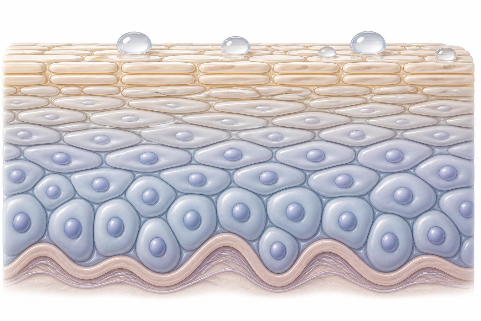
The keratinocyte is the most abundant cell in the epidermis, and it is the cell the world actually sees. Keratinocytes are born at the base of the epidermis and migrate upward over several weeks, maturing as they go, until they reach the surface as flattened, dead cells that form the stratum corneum — the true barrier of the skin.[28] This barrier does two jobs at once: it keeps water in, and it keeps irritants and pathogens out.[29]
The orderly maturation of the keratinocyte determines much of what patients mean when they talk about skin “quality.” When the process is healthy and the surface is well-organised, skin holds water, feels smooth, and reflects light evenly — the quality we perceive as “glow.” When the process is disordered — when dead cells accumulate unevenly, or the barrier is compromised — skin looks dull, feels rough, and loses water through a leaky surface, a phenomenon measured clinically as increased transepidermal water loss.[30]
Here the layered architecture returns to explain something practitioners observe daily. Because hydration diffuses upward from the dermis, a thick, disordered layer of dead surface cells impedes that water's passage and leaves the skin dry despite adequate moisture beneath.[4] This is why exfoliation and turnover-normalising treatments can make skin look and feel more hydrated without adding any water at all — they simply restore the surface's ability to receive what is already rising from below.
Treatments that target the keratinocyte are largely about regulating turnover and supporting the barrier. Chemical peels — using acids such as glycolic, salicylic or trichloroacetic acid at varying depths — accelerate the shedding of surface cells and prompt a more orderly regeneration of the epidermis.[31,32] Retinoids appear again here, because they normalise keratinocyte maturation as well as stimulating the fibroblast below — one of the reasons they are so uniquely valuable across the whole model.[11] Barrier-supporting topicals, containing ceramides, cholesterol and free fatty acids, help repair and maintain the stratum corneum itself.[33]
There is also a clinical caution that the keratinocyte teaches, which I have elsewhere termed “lazy skin”: the over-use of rich occlusive moisturisers can, in some patients, allow the skin's own barrier-maintenance machinery to become under-active, so that the skin becomes dependent on the external product.[34] The principle — that skin functions best when its own cells are kept working rather than replaced — recurs throughout this model.
6. Putting the three cells together
The value of the model appears when the three cells are considered as a system rather than in isolation, because the most powerful treatments act on more than one. The retinoid is the clearest example. It stimulates the fibroblast to build collagen, and it normalises the keratinocyte's maturation at the surface — two of the three cells addressed by a single, inexpensive, decades-old molecule.[11,12] This is precisely why it earns its place as the foundation of almost every serious skin-rejuvenation regime, and why a practitioner who understands the model reaches for it first.
Photoprotection is the second unifying intervention. Ultraviolet radiation is the principal external enemy of all three cells: it degrades the fibroblast's collagen, provokes the melanocyte into uneven pigment, and damages keratinocyte DNA.[9,20,27] Sunscreen is therefore not a cosmetic afterthought but the single most effective anti-ageing intervention available, because it protects the whole cellular system at once. The model makes this obvious in a way that a list of individual treatments never could.
The system view also imposes an order of operations. Because hydration and repair flow upward from the dermis, and because a provoked melanocyte can undermine any result, the rational sequence is to build the dermal foundation (fibroblast), respect and protect the pigment cell (melanocyte), and refine the surface last (keratinocyte) — while protecting all three from ultraviolet damage throughout. This biological sequencing is the subject of a companion article and is noted here only to show that the model generates a protocol, not merely a description.
7. Limitations
This model is a teaching device and should be understood as such. Skin biology is vastly more intricate than three cells; the immune system, the microbiome, the vasculature, hormonal signalling and the extracellular matrix all participate in ageing and repair in ways this framework deliberately omits.[2,35] Some treatments act through mechanisms that do not map neatly onto a single cell. The Fitzpatrick scale, invoked here for the melanocyte's reactivity, is itself an imperfect proxy for the biology of pigmentary risk.[36] None of this invalidates the model; it simply defines its scope. The Three-Cell Model is offered as a scaffold for understanding and for teaching, onto which the full complexity of the science can be added as the practitioner advances — not as a substitute for that complexity.
8. Conclusion
The practitioner who learns skin rejuvenation as a list of products will always be one launch behind the market. The practitioner who learns it as the behaviour of three cells — the fibroblast that builds, the melanocyte that must be respected, and the keratinocyte that faces the world — has a framework that does not go out of date.
When the next booster, device or acid appears, they need only ask which cell it acts on and how. That question, asked consistently, converts a bewildering marketplace into an ordered discipline, and it is the single most useful habit of thought I know how to teach.
9. Conflict of interest
The author is the founder and Medical Director of the Harley Street Institute, which provides commercial aesthetic training, and is the author of a forthcoming book on aesthetic practice. This manuscript was handled independently of the journal's Editor-in-Chief.
10. Funding
None.
AI Disclosure
Drafting and reference formatting were assisted by an AI language model under the author's direction and review. The author is responsible for all clinical content and conclusions.
Competing Interests
The author(s) declare no competing financial or non-financial interests relevant to this work.
Funding
This work received no specific grant from any funding agency in the public, commercial, or not-for-profit sectors.
Ethics & Consent
Where applicable, ethical approval and informed patient consent were obtained in accordance with the Declaration of Helsinki. Reviews and commentaries did not require ethical approval.
The Anatomy of Injection
A three-part framework linking cellular biology, tissue planes and injection technique into a single teaching model.
- 1/3The Three-Cell Model of Skin RejuvenationYou are here
- 2/3The Three-Layer Filler Model: Depth as the Anatomy of Injection
- 3/3The HSI Mini Scale: Biological Sequencing of Skin, Movement, Support and Descent
Companion reading
Where the pigment evidence for this framework sits
Pigment is assessed as skin quality, not structural support. The melanocyte component of this model is examined study by study in the companion evidence review.
- Journal · Systematic reviewClinical Efficacy and Safety of Pigment Modulators in Melasma and PIHThe evidence review behind the melanocyte component of this framework.
- White paperBeyond “Melanin Inhibitors”Direct enzyme-targeting agents versus indirect pigment modulators, and the language that follows from the distinction.
- Online bookAesthetic Talk — The EpidermisHow melanocyte behaviour and phototype risk are taught in the course.
References
- Sundaram H, Signorini M, Liew S, et al. Global Aesthetics Consensus: botulinum toxin type A — evidence-based review, emerging concepts, and consensus recommendations. Plast Reconstr Surg. 2016;137(3):518e–529e.
- Kolarsick PAJ, Kolarsick MA, Goodwin C. Anatomy and physiology of the skin. J Dermatol Nurses Assoc. 2011;3(4):203–213.
- Wong R, Geyer S, Weninger W, et al. The dynamic anatomy and patterning of skin. Exp Dermatol. 2016;25(2):92–98.
- Menon GK, Cleary GW, Lane ME. The structure and function of the stratum corneum. Int J Pharm. 2012;435(1):3–9.
- Frantz C, Stewart KM, Weaver VM. The extracellular matrix at a glance. J Cell Sci. 2010;123(24):4195–4200.
- Papakonstantinou E, Roth M, Karakiulakis G. Hyaluronic acid: a key molecule in skin aging. Dermatoendocrinol. 2012;4(3):253–258.
- Varani J, Dame MK, Rittie L, et al. Decreased collagen production in chronologically aged skin. Am J Pathol. 2006;168(6):1861–1868.
- Pittayapruek P, Meephansan J, Prapapan O, et al. Role of matrix metalloproteinases in photoaging and photocarcinogenesis. Int J Mol Sci. 2016;17(6):868.
- Gilchrest BA. Photoaging. J Invest Dermatol. 2013;133(E1):E2–E6.
- Fisher GJ, Kang S, Varani J, et al. Mechanisms of photoaging and chronological skin aging. Arch Dermatol. 2002;138(11):1462–1470.
- Mukherjee S, Date A, Patravale V, et al. Retinoids in the treatment of skin aging. Clin Interv Aging. 2006;1(4):327–348.
- Kang S, Fisher GJ, Voorhees JJ. Photoaging: pathogenesis, prevention, and treatment. Clin Geriatr Med. 2001;17(4):643–659.
- Hou A, Cohen B, Haimovic A, et al. Microneedling: a comprehensive review. Dermatol Surg. 2017;43(3):321–339.
- Alexiades-Armenakas MR, Dover JS, Arndt KA. The spectrum of laser skin resurfacing. J Am Acad Dermatol. 2008;58(5):719–737.
- Sadick N, Rothaus KO. Minimally invasive radiofrequency devices. Clin Plast Surg. 2016;43(3):567–575.
- Fitzgerald R, Vleggaar D. Using poly-L-lactic acid in facial rejuvenation. Facial Plast Surg. 2011;27(2):111–117.
- Kadouch JA. Calcium hydroxylapatite: a review on safety and complications. J Cosmet Dermatol. 2017;16(2):152–161.
- Cavallini M, Papagni M. Polynucleotides in aesthetic medicine: a review. J Cosmet Dermatol. 2023;22(7):1863–1869.
- Cichorek M, Wachulska M, Stasiewicz A, et al. Skin melanocytes: biology and development. Postepy Dermatol Alergol. 2013;30(1):30–41.
- Brenner M, Hearing VJ. The protective role of melanin against UV damage in human skin. Photochem Photobiol. 2008;84(3):539–549.
- Costin GE, Hearing VJ. Human skin pigmentation: melanocytes modulate skin color in response to stress. FASEB J. 2007;21(4):976–994.
- Passeron T, Picardo M. Melasma, a photoaging disorder. Pigment Cell Melanoma Res. 2018;31(4):461–465.
- Davis EC, Callender VD. Postinflammatory hyperpigmentation: a review of the epidemiology, clinical features, and treatment options in skin of color. J Clin Aesthet Dermatol. 2010;3(7):20–31.
- Alexis AF, Sergay AB, Taylor SC. Common dermatologic disorders in skin of color. Cutis. 2007;80(5):387–394.
- Pillaiyar T, Manickam M, Namasivayam V. Skin whitening agents: medicinal chemistry perspective of tyrosinase inhibitors. J Enzyme Inhib Med Chem. 2017;32(1):403–425.
- Sarkar R, Arora P, Garg VK, et al. Melasma update. Indian Dermatol Online J. 2014;5(4):426–435.
- Randhawa M, Wang S, Leyden JJ, et al. Daily use of a broad-spectrum sunscreen over 1 year significantly improves photoaged skin. Dermatol Surg. 2016;42(12):1354–1361.
- Eckhart L, Lippens S, Tschachler E, et al. Cell death by cornification. Biochim Biophys Acta. 2013;1833(12):3471–3480.
- Proksch E, Brandner JM, Jensen JM. The skin: an indispensable barrier. Exp Dermatol. 2008;17(12):1063–1072.
- Akdeniz M, Gabriel S, Lichterfeld-Kottner A, et al. Transepidermal water loss in healthy adults: a systematic review. Br J Dermatol. 2018;179(5):1049–1055.
- Soleymani T, Lanoue J, Rahman Z. A practical approach to chemical peels. J Clin Aesthet Dermatol. 2018;11(8):21–28.
- Rendon MI, Berson DS, Cohen JL, et al. Evidence and considerations in the application of chemical peels. J Clin Aesthet Dermatol. 2010;3(7):32–43.
- Spada F, Barnes TM, Greive KA. Skin hydration is significantly increased by a cream formulated to mimic the skin's own natural moisturizing systems. Clin Cosmet Investig Dermatol. 2018;11:491–497.
- Rawlings AV, Harding CR. Moisturization and skin barrier function. Dermatol Ther. 2004;17(Suppl 1):43–48.
- Byrd AL, Belkaid Y, Segre JA. The human skin microbiome. Nat Rev Microbiol. 2018;16(3):143–155.
- Ware OR, Dawson JE, Shinohara MM, et al. Racial limitations of Fitzpatrick skin type. Cutis. 2020;105(2):77–80.
Declarations
- Peer review:
- This article underwent single-blind external peer review by at least two independent reviewers, followed by editorial acceptance.
- Conflicts of interest:
- The author(s) declare no competing financial or commercial interests relating to the content of this article. Editorial decisions are made independently of the Harley Street Institute's commercial training activities.
- Funding:
- No external funding was received for the preparation of this article.
- Licence:
- © 2026 Harley Street Institute. Open access article distributed under the Creative Commons Attribution 4.0 International Licence (CC BY 4.0), permitting unrestricted use with appropriate citation.
Post-publication review
Discuss this article with the journal AI
Ask a clinical question about this article, or flag a possible error. Our AI agent will reply in real time, log your input, and — if you have identified a credible mistake — escalate it to the HSI editorial team for review and a published correction notice.
AI responses are generated by an assistant model. They do not constitute medical advice. Editorial corrections are only applied after a named HSI editor reviews and signs off.
Hi. I'm the Aesthetic Intelligence reader AI for this article.
Try: "Explain section 3 in plain English." · "What's the evidence for the 1-in-6,410 figure?" · "How does this compare to UK NICE guidance?"