
Review Article · Aesthetic Intelligence · Vol 1 · Issue 7
The HSI Mini ScaleFour Domains, One Dominant Driver
Facial ageing is several clocks running at once, not one cascade from bone to skin. Score skin, movement, support and descent separately — then treat the domain that is actually producing the complaint.
Dr Ahmed Haq1
- 1 Founder and Medical Director, Harley Street Institute, London, United Kingdom
Corresponding author: journal@harleystreetinstitute.com
Journal: Aesthet Intell
DOI: to be assigned
Volume / Issue: 1 / 7
Pages: 35–48
Received: 2026-07-02
Accepted: 2026-07-08
Published: 2026-07-10
Licence: CC BY 4.0
Part three · The Anatomy of Injection
There is a comforting story told in aesthetic medicine, usually with a diagram: the bone goes first, then the fat slides south, then the skin gives up, and the injector arrives with a syringe to reverse the order of events. It is a tidy story. It is also, for most of the faces in front of us, wrong in the only way that matters — it tells you what happened to a population, not what is happening to this patient.
A twenty-eight-year-old with pigment and a rough surface does not need a periosteal bolus because bone resorption is somewhere in her future. A fifty-five-year-old with a heavy jawline does not need more volume simply because she is fifty-five. What both need is an examination that separates the face into things that can actually be looked at, one at a time, and a clinician willing to write down which one is doing the damage.
That is all the HSI Mini Scale is: four questions asked in a fixed order, scored honestly, and reported as a shape rather than a number.
Abstract
- Background.
- Facial ageing is frequently taught as a single chronological cascade in which bone resorbs, fat descends and skin fails, with treatment following the same deep-to-superficial order. Existing clinical scales largely measure one feature at a time — lines, sagging, volume or texture — and validation efforts have historically focused on grading one feature at a time against standardised photographs. Neither the cascade model nor a single-feature score tells the clinician which tissue is actually producing the patient's complaint.
- Methods.
- Narrative review and educational synthesis of published facial ageing scales, imaging studies of facial fat compartments, surface electromyography of facial musculature and consensus assessment recommendations, used to construct and define a four-domain examination framework — the proposed HSI Mini Scale — with defined findings, examination method and treatment families for each domain.
- Results.
- The framework separates the face into four assessment domains rather than four chronological stages: skin biology and quality; muscle and movement; volume, shape and support; and laxity and tissue descent. Each is scored 0–3 and reported as a phenotype (for example S2–M3–V0–D1) rather than summed, because identical totals describe clinically dissimilar patients. Two free-text fields — the patient's stated priority and the clinician-identified dominant driver — are mandatory. The examination sequence is fixed; the treatment sequence is determined by the dominant driver and not by anatomical depth.
- Conclusion.
- Presented as a proposed, not validated, instrument, the HSI Mini Scale operationalises the principle that anatomical ageing and treatment target are not the same thing. Formal validation would require photographic anchors, diverse patient representation, and inter- and intra-rater reliability testing against established domain scales.
Keywords: facial ageing assessment, aesthetic scales, skin quality, facial muscle activity, facial fat compartments, facial laxity, treatment sequencing, HSI Mini Scale
| Evidence domain | What the article reports | How to interpret it |
|---|---|---|
| Background | Facial ageing is frequently taught as a single chronological cascade in which bone resorbs, fat descends and skin fails, with treatment following the same deep-to-superficial order. Existing clinical scales largely measure one feature at a time — lines, sagging, volume or texture — and validation efforts have historically focused on grading one feature at a time against standardised photographs. Neither the cascade model nor a single-feature score tells the clinician which tissue is actually producing the patient's complaint. | Defines the problem and the reason for review or investigation. |
| Methods | Narrative review and educational synthesis of published facial ageing scales, imaging studies of facial fat compartments, surface electromyography of facial musculature and consensus assessment recommendations, used to construct and define a four-domain examination framework — the proposed HSI Mini Scale — with defined findings, examination method and treatment families for each domain. | Shows how the evidence or observations were assembled. |
| Results | The framework separates the face into four assessment domains rather than four chronological stages: skin biology and quality; muscle and movement; volume, shape and support; and laxity and tissue descent. Each is scored 0–3 and reported as a phenotype (for example S2–M3–V0–D1) rather than summed, because identical totals describe clinically dissimilar patients. Two free-text fields — the patient's stated priority and the clinician-identified dominant driver — are mandatory. The examination sequence is fixed; the treatment sequence is determined by the dominant driver and not by anatomical depth. | Summarises the principal findings reported by the authors. |
| Conclusion | Presented as a proposed, not validated, instrument, the HSI Mini Scale operationalises the principle that anatomical ageing and treatment target are not the same thing. Formal validation would require photographic anchors, diverse patient representation, and inter- and intra-rater reliability testing against established domain scales. | States the authors’ clinical interpretation and its limits. |
Postgraduate Level
Complete this article to earn your certificate
Read the article, complete a short assessment, and submit your reflection to receive your AiCE Points certificate. Equivalence to 1 CPD/CME point — we do not award CPD/CME directly.
Learning Objectives
- 1Assess the face as four examination domains rather than four chronological stages of ageing
- 2Distinguish dermal, dynamic, folding and contour causes of an apparent line
- 3Recognise that facial fat may decrease, increase, redistribute or descend, and avoid assuming deficit
- 4Differentiate cutaneous laxity, true soft-tissue descent, pseudoptosis and apparent sagging from excess
- 5Report an S–M–V–D phenotype with a dominant driver and state what is not currently indicated
The HSI Mini Scale at a glance
Four domains
- S — Skin Quality
- Barrier, pigment, matrix, vascular modifiers
- M — Muscle & Movement
- Recruitment, tone, balance, asymmetry
- V — Volume & Support
- Bone, projection, fat, previous filler
- D — Laxity & Descent
- Recoil, displacement, pseudoptosis, excess
Scoring
- 0
- No meaningful finding
- 1
- Early, mild or localised
- 2
- Clearly established or moderate
- 3
- Advanced, marked or multifocal
- Report as
- Phenotype (e.g. S2–M3–V0–D1) — never a total
Mandatory fields
- Patient priority
- The concern in the patient's own words
- Dominant driver
- The tissue or mechanism producing it
1. Introduction: several clocks, not one cascade
Facial ageing is commonly taught as a sequence. Bone resorbs, deep fat deflates, retaining structures weaken, superficial tissue descends and the skin — arriving last — merely records the result. The teaching is memorable, and its clinical corollary is equally memorable: treat deep first, superficial last. The difficulty is that the cascade describes an average trajectory across populations, whereas the clinician treats one face, on one day, with one dominant complaint.
The measurement literature reflects the same fragmentation. Published clinical ageing scales overwhelmingly assess a single feature — lines and wrinkles, sagging or volume, texture and skin quality — with a smaller number offering global impressions; the wrinkle severity rating scale, for example, was validated as a single-feature photonumeric instrument.[1] Glogau's classification, one of the earliest widely used systems, already separated wrinkles at rest from wrinkles in motion, implicitly conceding that a face contains more than one process.[2] Later validated photonumeric scales followed the same logic, grading upper- and lower-face features individually and against standardised photographs.[3,4]
What is missing is not another wrinkle score or another jawline grade. It is a single, short, causal assessment that asks which tissue is producing the patient's concern before any product is selected. That is the purpose of the proposed HSI Mini Scale. It is the third part of a series: the first proposed a three-cell model of skin biology,[12] the second proposed that, for the injector, anatomy is depth.[13] This paper supplies the missing step between them — the examination that decides whether either model should be used at all.
One clarification governs everything that follows. The four elements of the scale are domains of examination, not stages of ageing. No patient is required to pass through them in order, and a high score in one domain does not oblige the clinician to expect or pre-empt the others.
2. The HSI Mini Scale: a four-domain examination
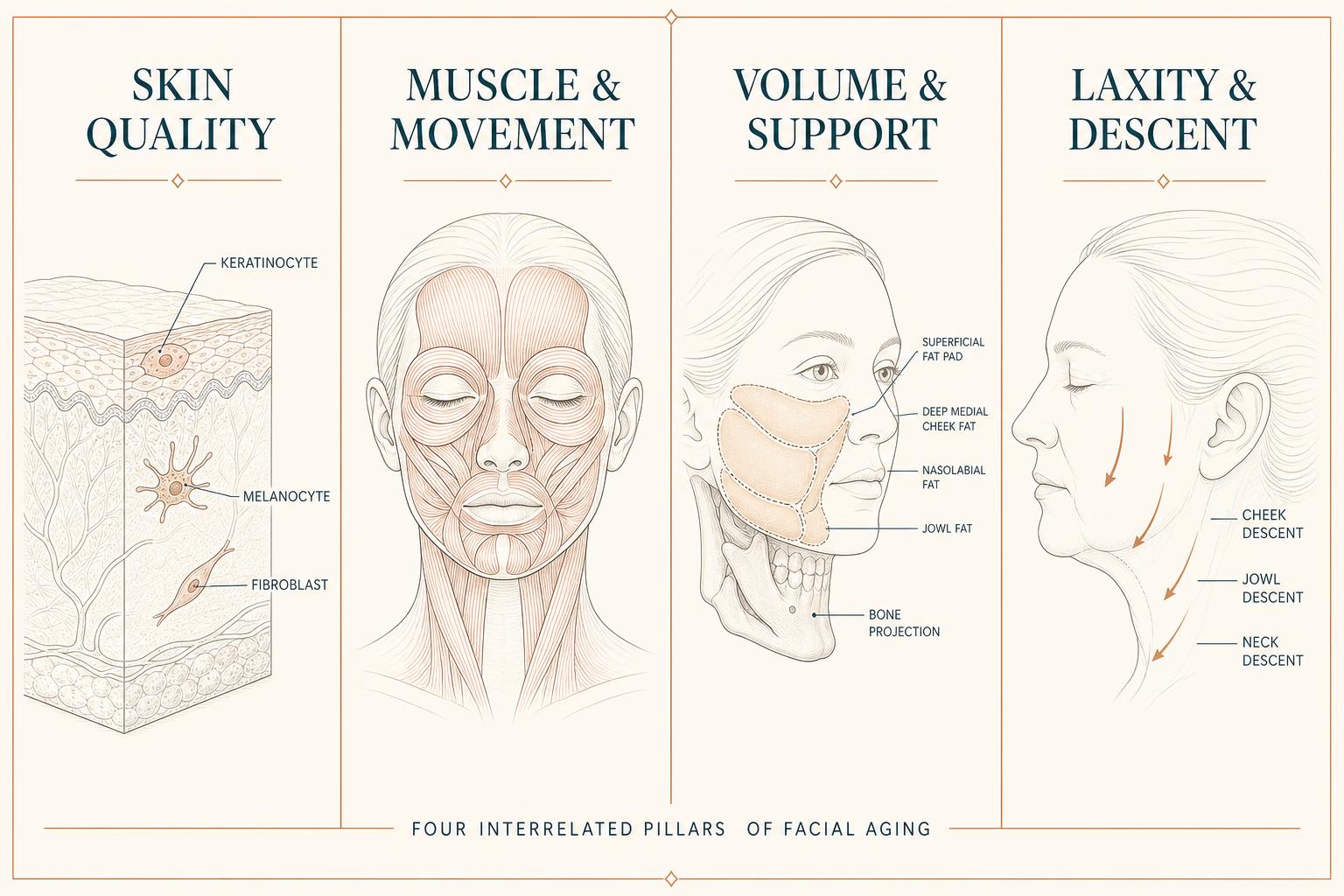
The scale comprises four domains: skin biology and quality; muscle and movement; volume, shape and support; and laxity and tissue descent. In the clinical interface these appear as four cards — Skin Quality, Muscle & Movement, Volume & Support, Laxity & Descent.
The wording matters. Three commonly used shorthand terms have been deliberately rejected. “Wrinkles in motion” is too narrow, because it omits resting tone, asymmetry, hypertrophy, atrophy and the balance between opposing muscle groups. “Volume loss” presumes a deficit, whereas facial fat may decrease, increase, redistribute or descend, and a patient may be congenitally underprojected or carrying excessive previous filler. “Sagging skin” implies that the skin alone has fallen, when apparent sagging may involve dermis, superficial fat, retaining structures, superficial musculoaponeurotic system, muscle, bone, volume excess and gravity in varying proportion.
The novelty claimed here is modest and specific: not a new measurement of any one feature, but the combination of four established domains into one short causal assessment whose output is a treatment decision rather than a severity grade.
3. Domain one — Skin quality
The question is simple: is the patient's principal concern located within the skin itself?
The domain maps directly onto the three-cell model.[12] Keratinocyte-related findings include impaired barrier function, dryness, roughness, dullness, uneven texture, superficial scaling, reduced epidermal resilience and fine surface lines. Melanocyte-related findings include lentigines, mottled pigmentation, post-inflammatory hyperpigmentation, melasma-pattern pigmentation, uneven tone and hypopigmented areas; risk is strongly modified by Fitzpatrick phototype.[11] Fibroblast and extracellular-matrix findings include fine static lines, crepiness, dermal thinning, reduced recoil, loss of firmness, textural irregularity and atrophic scarring. Vascular and inflammatory modifiers — erythema, telangiectasia, rosacea-pattern inflammation and chronic irritation — are recorded here even though they sit outside the original three-cell teaching model.
Examination is performed without make-up, at rest, in diffuse frontal light and again in oblique light to reveal surface irregularity, with attention to pigment, erythema, pores, texture, hydration, scarring and fine lines, and with gentle palpation or a recoil assessment where appropriate. The examiner must distinguish a line caused principally by matrix damage from a line produced by muscle movement, a line created by folding or descent, and a shadow created by contour.
Treatment families include photoprotection, barrier repair, retinoids and other evidence-based topical therapy, pigment-modulating treatment, chemical peeling, microneedling, fractional resurfacing, vascular- or pigment-directed devices, radiofrequency-based dermal treatment and selected collagen-stimulating or regenerative approaches. Selection depends on phototype, diagnosis, treatment history, pigmentary risk and local regulatory approval.
The central warning of this domain is that a skin-quality problem should not automatically receive filler. A younger patient scoring S2–M1–V0–D0 may require an entirely superficial sequence — protect, regulate, then regenerate or resurface — with no structural treatment whatsoever, because deeper anatomical change that may occur later is not a present indication.
4. Domain two — Muscle and movement
The question is broader than dynamic lines: what happens when the face moves, and is muscle activity the dominant driver of the patient's concern?
The assessment records dynamic recruitment, resting tone, relative strength, elevator–depressor balance, asymmetry, compensatory activity, hypertrophy, atrophy and the transition from a dynamic line to a static dermal imprint. Muscles should not be assumed to weaken uniformly with age; surface electromyography studies have not demonstrated a single global decline in facial muscle signal with age, with differences instead appearing in specific muscles such as zygomaticus major, procerus and corrugator supercilii.[5] Muscle behaviour is therefore regional and patient-specific.
A standardised movement sequence is proposed: face fully at rest; brow elevation; frowning; gentle and then maximal eye closure; squinting; gentle smile; full smile; nasal scrunch; lip pursing; oral-commissure depression; chin contraction; and platysmal contraction where relevant. Consensus recommendations for upper-face treatment already emphasise visual and tactile assessment at rest and during animation, together with evaluation of opposing-muscle interaction.[14]
A finding may legitimately belong to more than one domain. A lateral canthal line visible only on maximal smiling is predominantly a movement finding; one that persists at rest is both movement and skin; a deeply etched glabellar line may require movement control first and reassessment of the dermal component afterwards. A nasolabial fold is not a muscle problem merely because it deepens on smiling. Validated scales have long graded wrinkles at rest and on movement separately for exactly this reason.[3,4]
Treatment families include selective neuromodulation of demonstrably overactive muscles, rebalancing of opposing groups, deliberate avoidance of already weak or compensating muscles, reassessment after modulation, skin-directed treatment of the residual static imprint and, very selectively, structural treatment of a persistent crease. The objective is not the abolition of movement but the modulation of excessive or unbalanced movement with expression preserved.
5. Domain three — Volume, shape and support
This domain is deliberately titled volume, shape and support rather than volume loss, because not every depression or shadow represents missing tissue. It encompasses the facial skeleton and projection, dental and maxillary support, deep and superficial fat compartments, temporal and periorbital tissue, buccal fat, previous filler, weight-related change, congenital proportion, localised lipoatrophy and muscle bulk where it contributes to shape.[8]
Imaging does not support uniform facial fat loss. Longitudinal computed tomography has reported an increase in deep cheek fat volume over roughly a decade,[6] while magnetic resonance studies have reported significant reductions in superficial and deep midfacial fat over comparable intervals,[7] and three-dimensional analyses have attributed much of apparent midfacial ageing to descent and redistribution rather than simple deflation.[9] These findings are not contradictory so much as regional, method-dependent and patient-specific. Skeletal change compounds the picture: maxillary and orbital remodelling alters the platform on which the soft tissue sits.[10]
The examiner therefore asks whether volume is genuinely absent; whether tissue has changed position; whether the patient is congenitally underprojected; whether skeletal or dental support is deficient; whether the shadow is created by skin or by a ligamentous attachment; whether there is in fact volume excess; whether previous filler has altered the anatomy; whether body weight has changed; and whether the finding persists during expression.
Assessment is made in frontal, three-quarter and profile views, with a basal or submental view where relevant, in neutral expression and on smiling, upright, with palpation where useful, and with reference to previous photographs, treatment history, dental history and any substantial weight change.
Treatment families include small localised hyaluronic-acid correction, structural projection at a carefully selected point, subcutaneous contour correction, fat grafting, selected biostimulatory treatment, correction or dissolution of inappropriate previous filler, dental or orthognathic assessment, and implant or surgical options in selected patients. The scale must never generate the conclusion that a patient is older and therefore requires volume replacement. The correct question is whether an identifiable deficit is materially contributing to the stated concern, and whether correcting it would improve the face without making it heavier or unfamiliar.
6. Domain four — Laxity and tissue descent
Renaming this domain from “sagging skin” to laxity and tissue descent is the most consequential change in the framework, because apparent sagging is a multilayer phenotype. Contributors include reduced dermal elasticity, superficial fat displacement, change in fibrous septa and retaining structures, alteration in the superficial musculoaponeurotic system and fascia, muscle change, platysmal activity, reduced structural projection, submental or lower-face volume excess, weight fluctuation, gravity and previous overfilling. Quantitative work has associated perceived sagging with a combination of dermal viscoelasticity, subcutaneous fat thickness and muscle thickness, with the relative contribution differing by region and age.[7]
Four presentations must be distinguished. Cutaneous laxity: reduced recoil, crepiness or looseness with limited underlying descent. Soft-tissue descent: displaced superficial tissue producing malar flattening, nasolabial deepening, jowling or interruption of the mandibular outline. Pseudoptosis from deflation or underprojection: tissue appears to hang because support is inadequate. Apparent sagging from excess: lower-face heaviness, fat excess or excessive previous filler creating or worsening a descended appearance. These four cannot receive the same treatment.
Examination includes skin recoil and mobility, pinch characteristics, jowl formation, mandibular-border interruption, malar position, nasolabial and marionette configuration, preauricular laxity, submental and cervical tissue, platysmal banding, comparison at rest and on expression, and gentle manual redraping. Manual repositioning helps explain the phenotype; it does not demonstrate that a filler-based lift is appropriate.
Treatment families include skin-quality optimisation, collagen-stimulating approaches, selected energy-based tightening, limited support correction where a genuine deficit coexists, reduction or correction of excess volume, thread-based or minimally invasive approaches in carefully selected cases, surgical lifting for established soft-tissue ptosis, and no treatment where intervention would make the face heavier. Filler is not a substitute for tissue repositioning; adding volume to an already heavy or descended lower face increases distortion rather than producing a lift.
7. Scoring: a phenotype, not a total
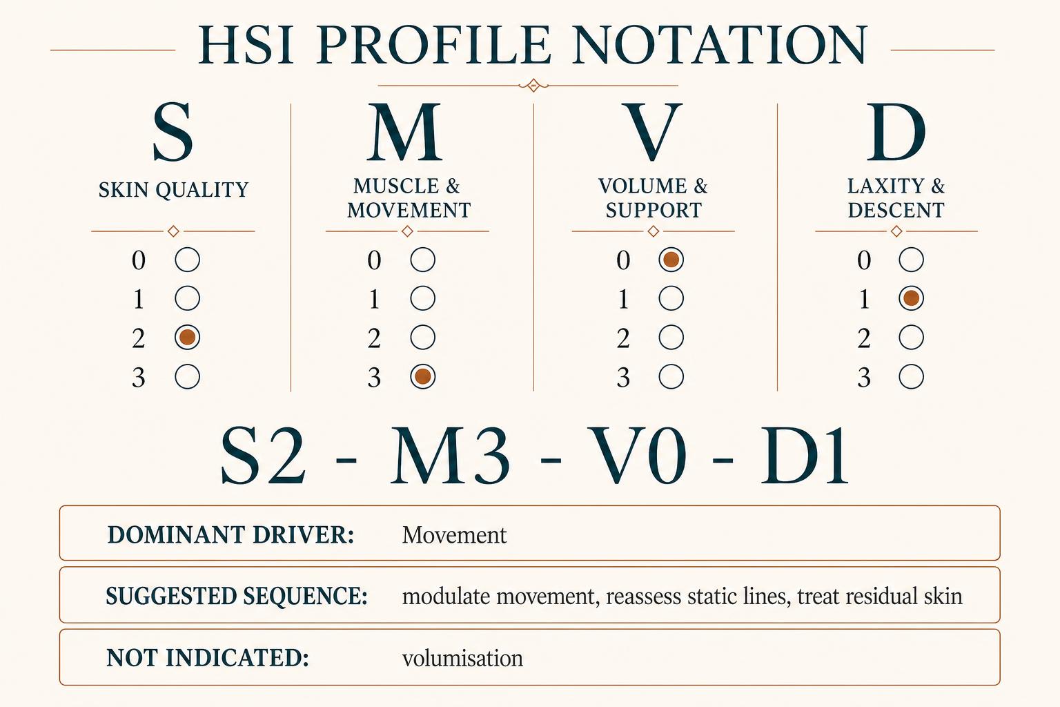
Each domain is scored 0–3. A score of 0 indicates no meaningful finding; 1 an early, mild or localised finding; 2 a clearly established or moderate finding; and 3 an advanced, marked or multifocal finding.
The four scores are not summed. A total of six could represent severe skin disease with no structural change, advanced descent with otherwise excellent skin, or moderate abnormality across three domains — patients who are not clinically equivalent and should not share a label. The result is therefore reported as a phenotype: for example, HSI profile S2–M3–V0–D1, meaning moderate skin findings, advanced movement findings, no meaningful volume or support deficit and mild descent.
The output displayed to the clinician is correspondingly causal rather than numerical: dominant driver (movement); suggested sequence (modulate movement, reassess static lines, treat the residual skin component); not currently indicated (volumisation). This is safer and more educational than reporting “score 6: moderate ageing”, and it prevents the scale from being read as a biological-age estimate.
8. Two fields the numbers cannot capture
Four severity scores are insufficient on their own. Two free-text fields are mandatory.
The first is the patient priority: what is actually bothering the patient, recorded in their words — for example, “lines around my eyes when I smile”. The second is the clinician-identified dominant driver: the tissue or mechanism contributing most to that specific concern — for example, “strong lateral orbicularis activity with a mild residual dermal imprint”.
These are not always the same, and recording both prevents the instrument from degenerating into a mechanical treatment calculator that treats every scored abnormality it detects.
9. The examination sequence can be standardised; the treatment sequence cannot
The consultation follows a fixed order. Observe at rest, assessing skin, pigmentation, static lines, shape, shadow, symmetry, contour and descent. Observe in motion, assessing recruitment, muscle balance, line formation, tissue movement and expression. Assess shape and support, examining projection, fat distribution, contour, structural transitions, previous filler and relevant history. Assess laxity and descent, distinguishing dermal laxity, soft-tissue displacement, structural underprojection and lower-face heaviness. Identify the dominant driver. Choose the least invasive effective target — and do not treat every scored abnormality.
Treatment order, by contrast, follows the dominant driver. In a skin-dominant patient: protect, regulate, regenerate or resurface, then reassess; there is no requirement to inject the deep face first. In a movement-dominant patient: modulate movement, allow the result to stabilise, reassess the static component, and treat residual skin or structural change only where necessary. In a volume- or support-dominant patient: correct the smallest meaningful deficit, reassess the visible fold or shadow, and refine only if needed. In a descent-dominant patient: determine whether the problem is mild laxity, true ptosis, excess volume or lost support, establish the non-surgical ceiling, and tighten, reposition or refer appropriately. This last pathway prevents the familiar error of attempting to correct descent with repeated volumisation.
10. Worked examples
Young patient with pigmentation and fine texture — profile S2–M1–V0–D0. Driver: skin. Sequence: photoprotection and skin-directed treatment. Avoid: prophylactic cheek or periosteal filler.
Young patient with strong glabellar activity — profile S1–M3–V0–D0. Driver: muscle activity. Sequence: selective neuromodulation, then reassessment. Avoid: filling the line before controlling the repetitive mechanical driver.
Patient with a genuine localised support deficit — profile S1–M1–V2–D1. Driver: volume and support. Sequence: conservative structural or contour correction, then reassessment of the overlying fold. Avoid: filling every visible line or replacing all presumed age-related loss.
Patient with heavy jowls and advanced descent — profile S1–M1–V0–D3. Driver: descent. Sequence: establish the surgical versus non-surgical ceiling. Avoid: attempting to lift the lower face by increasing filler volume.
Patient with platysmal banding and cervical laxity — profile S2–M2–V0–D2. Drivers: movement and descent. Sequence: consider muscle modulation and laxity treatment as separate but related targets. Avoid: treating the neck as a volume-loss problem.
11. Worked case: an itemised before-and-after assessment
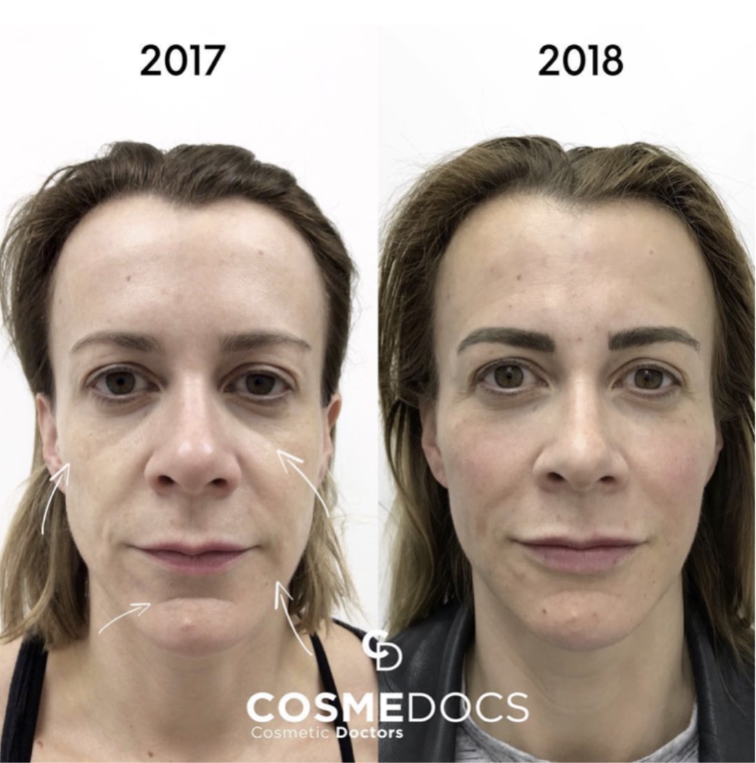
The domain profile is the clinical output of the scale, but teaching and audit often require the underlying item scores to be written down. The itemised form below — the HSI Mini Facial Scale (antiageing) — records the same four domains at item level, scored 0–3 for surface, volume and laxity items and 0–4 for static lines, before treatment and again at review. The client had received previous upper-face botulinum toxin, so the static-line domain was already controlled at baseline.
Skin surface, before then after: pores size and congestion 1 / 1; colour changes 1 / 1; hydration 1 / 1; roughness 1 / 1; sensitivity 0 / 0.
Static lines, before then after: frown 0 / 0; forehead 0 / 0; crow's feet 0 / 0; DAO 0 / 0; bunny lines 0 / 0; oral commissure 1 / 0; mentalis crease 3 / 1.
Volume and structure, before then after: temples 1 / 0; cheeks 2 / 1; nasolabial 2 / 0; chin 2 / 0; jawline 3 / 1; marionette 2 / 1; lips 1 / 1; tear trough 1 / 1.
Skin laxity, before then after: jawline laxity 1 / 1; eyebrow hooding not scored.
Totals fell from 23 to 11. The instructive detail is not the fall in the total but its distribution: the entire change sits in the volume, support and lower-face line items, while skin-surface and laxity items are unchanged. The dominant driver was correctly identified as structural, treatment was sequenced accordingly, and no skin-directed or lifting procedure was performed for findings that were never contributing to the complaint. Item totals are used here descriptively for a single patient over time; they are not summed across domains to generate a severity grade, for the reasons set out in section 7.
A blank version of this form, with empty Before and After columns, accompanies the online article for clinicians who wish to score their own patients.
| HSI Mini Facial Scale — Antiageing (worked example) | |||
|---|---|---|---|
| Item | Before | After | Change |
| Skin Surface (0,1,2,3) | |||
| Pores Size & Congestion | 1 | 1 | 0 |
| Colour Changes | 1 | 1 | 0 |
| Hydration | 1 | 1 | 0 |
| Roughness | 1 | 1 | 0 |
| Sensitivity | 0 | 0 | 0 |
| Static Lines (0,1,2,3,4) | |||
| Frown | 0 | 0 | 0 |
| Forehead | 0 | 0 | 0 |
| Crows Feet | 0 | 0 | 0 |
| DAO (Sad Expression Lines) | 0 | 0 | 0 |
| Bunny Lines | 0 | 0 | 0 |
| Oral Commissure | 1 | 0 | -1 |
| Mentalis Crease | 3 | 1 | -2 |
| Volume & Structure (0,1,2,3) | |||
| Temples | 1 | 0 | -1 |
| Cheeks | 2 | 1 | -1 |
| Nasolabial | 2 | 0 | -2 |
| Chin | 2 | 0 | -2 |
| Jawline | 3 | 1 | -2 |
| Marionette | 2 | 1 | -1 |
| Lips | 1 | 1 | 0 |
| Tear Trough | 1 | 1 | 0 |
| Skin Laxity (0,1,2,3) | |||
| Jawline Laxity | 1 | 1 | 0 |
| Eyebrow Hooding | — | ||
| Total | 23 | 11 | -12 |
Client had previous upper face botulinum toxin, so the static-line domain is already controlled at baseline.
Use this scale in your own consultations — download the same form with empty Before and After columns and score your own patient.
12. Status, validation requirements and limitations
The instrument is presented as the proposed HSI Mini Scale. It has not been validated, and it should not be described as validated in teaching, marketing or clinical documentation until it has been.
Validation would require photographic anchors for each score in each domain, representation of diverse phototypes, ages and facial morphologies, repeated ratings by multiple assessors, formal inter- and intra-rater reliability testing, comparison against established single-domain scales, and demonstration that scores respond consistently to clinical change. Existing validated aesthetic scales have been developed in precisely this way, using standardised photographic references and repeated expert rating.[3,4]
Two further limitations are acknowledged. The framework is a narrative synthesis, not a systematic review, and the imaging literature it draws on is heterogeneous in method and cohort. And the scale is an examination aid: it structures observation and constrains over-treatment, but it does not replace diagnosis, consent, or supervised anatomical and practical training.
13. Conclusion
Facial ageing is the superimposition of several tissue clocks rather than a single cascade from bone to skin. Biological sequencing does not mean treating every face from deep to superficial; it means examining the relevant biological domains, identifying the dominant driver and choosing the least invasive effective target.
Three sequences should therefore be kept distinct. The biological sequence describes what is changing in skin, muscle, fat, retaining structures and bone, and over what timescale. The examination sequence is fixed: skin, then movement, then volume and support, then laxity and descent. The treatment sequence is determined by the dominant driver, followed by reassessment and residual refinement.
Held apart, these three sequences protect the reader from a series of persistent assumptions: that bone always ages first, that skin always ages last, that deep treatment must precede superficial treatment, that every volume change is volume loss, that every jowl requires filler, that every dynamic line requires toxin, and that every age-related anatomical change requires correction. Anatomical ageing and treatment target are not the same thing, and the examination exists to tell them apart.
AI Disclosure
Generative AI tools were used for language editing, reference formatting and figure drafting. All clinical content, framework design, interpretation and final wording are the author's own, and the author accepts full responsibility for the published text.
Competing Interests
The author(s) declare no competing financial or non-financial interests relevant to this work.
Funding
This work received no specific grant from any funding agency in the public, commercial, or not-for-profit sectors.
Ethics & Consent
Where applicable, ethical approval and informed patient consent were obtained in accordance with the Declaration of Helsinki. Reviews and commentaries did not require ethical approval.
HSI Editorial · Reflection & Forward Recommendations
Where we stand on this
Reflection
The commonest error in aesthetic consultation is not poor technique but premature treatment selection — choosing the product before naming the problem.
A scale that produces a single ageing score invites exactly that error. A scale that produces a phenotype and a dominant driver makes it harder to commit.
Forward Recommendations
- Record the four domain scores and the patient's stated priority in every consultation note.
- State explicitly what is not currently indicated — it is as clinically useful as what is.
- Reassess after treating the dominant driver before adding a second modality.
- Describe the instrument as the proposed HSI Mini Scale until validation data exist.
Editorial position of the Harley Street Institute. Authored by the HSI Clinical Review Board; not a substitute for the peer-reviewed evidence summarised above.
The Anatomy of Injection
A three-part framework linking cellular biology, tissue planes and injection technique into a single teaching model.
- 1/3The Three-Cell Model of Skin Rejuvenation
- 2/3The Three-Layer Filler Model: Depth as the Anatomy of Injection
- 3/3The HSI Mini Scale: Biological Sequencing of Skin, Movement, Support and DescentYou are here
References
- Day DJ, Littler CM, Swift RW, Gottlieb S. The wrinkle severity rating scale: a validation study. Am J Clin Dermatol. 2004;5(1):49-52.
- Glogau RG. Aesthetic and anatomic analysis of the aging skin. Semin Cutan Med Surg. 1996;15(3):134-8.
- Flynn TC, Carruthers A, Carruthers J, et al. Validated assessment scales for the upper face. Dermatol Surg. 2012;38(2 Pt 2):309-19.
- Narins RS, Carruthers J, Flynn TC, et al. Validated assessment scales for the lower face. Dermatol Surg. 2012;38(2 Pt 2):333-42.
- Schumann NP, Bongers K, Scholle HC, Guntinas-Lichius O. Facial muscle activation patterns in healthy male humans: a multi-channel surface EMG study. J Neurosci Methods. 2010;187(1):120-8.
- Gierloff M, Stöhring C, Buder T, Gassling V, Açil Y, Wiltfang J. Aging changes of the midfacial fat compartments: a computed tomographic study. Plast Reconstr Surg. 2012;129(1):263-73.
- Wysong A, Joseph T, Kim D, Tang JY, Gladstone HB. Quantifying soft tissue loss in facial aging: a study in women using magnetic resonance imaging. Dermatol Surg. 2013;39(12):1895-902.
- Rohrich RJ, Pessa JE. The fat compartments of the face: anatomy and clinical implications for cosmetic surgery. Plast Reconstr Surg. 2007;119(7):2219-27.
- Coleman SR, Grover R. The anatomy of the aging face: volume loss and changes in 3-dimensional topography. Aesthet Surg J. 2006;26(1S):S4-9.
- Mendelson B, Wong CH. Changes in the facial skeleton with aging: implications and clinical applications in facial rejuvenation. Aesthetic Plast Surg. 2012;36(4):753-60.
- Fitzpatrick TB. The validity and practicality of sun-reactive skin types I through VI. Arch Dermatol. 1988;124(6):869-71.
- Haq A. The Three-Cell Model of skin rejuvenation: a simplified cellular framework for aesthetic practice. Aesthet Intell. 2026;1(7).
- Haq A. The Three-Layer Filler Model: depth as the anatomy of injection. Aesthet Intell. 2026;1(7).
- Sundaram H, Signorini M, Liew S, et al. Global aesthetics consensus: botulinum toxin type A — evidence-based review, emerging concepts, and consensus recommendations for aesthetic use, including updates on complications. Plast Reconstr Surg. 2016;137(3):518e-529e.
Declarations
- Peer review:
- This article underwent single-blind external peer review by at least two independent reviewers, followed by editorial acceptance.
- Conflicts of interest:
- The author(s) declare no competing financial or commercial interests relating to the content of this article. Editorial decisions are made independently of the Harley Street Institute's commercial training activities.
- Funding:
- No external funding was received for the preparation of this article.
- Licence:
- © 2026 Harley Street Institute. Open access article distributed under the Creative Commons Attribution 4.0 International Licence (CC BY 4.0), permitting unrestricted use with appropriate citation.
Post-publication review
Discuss this article with the journal AI
Ask a clinical question about this article, or flag a possible error. Our AI agent will reply in real time, log your input, and — if you have identified a credible mistake — escalate it to the HSI editorial team for review and a published correction notice.
AI responses are generated by an assistant model. They do not constitute medical advice. Editorial corrections are only applied after a named HSI editor reviews and signs off.
Hi. I'm the Aesthetic Intelligence reader AI for this article.
Try: "Explain section 3 in plain English." · "What's the evidence for the 1-in-6,410 figure?" · "How does this compare to UK NICE guidance?"