Review Article · Aesthetic Intelligence · Vol 1 · Issue 7
The Three-Layer Filler ModelDepth Is the Anatomy of Injection
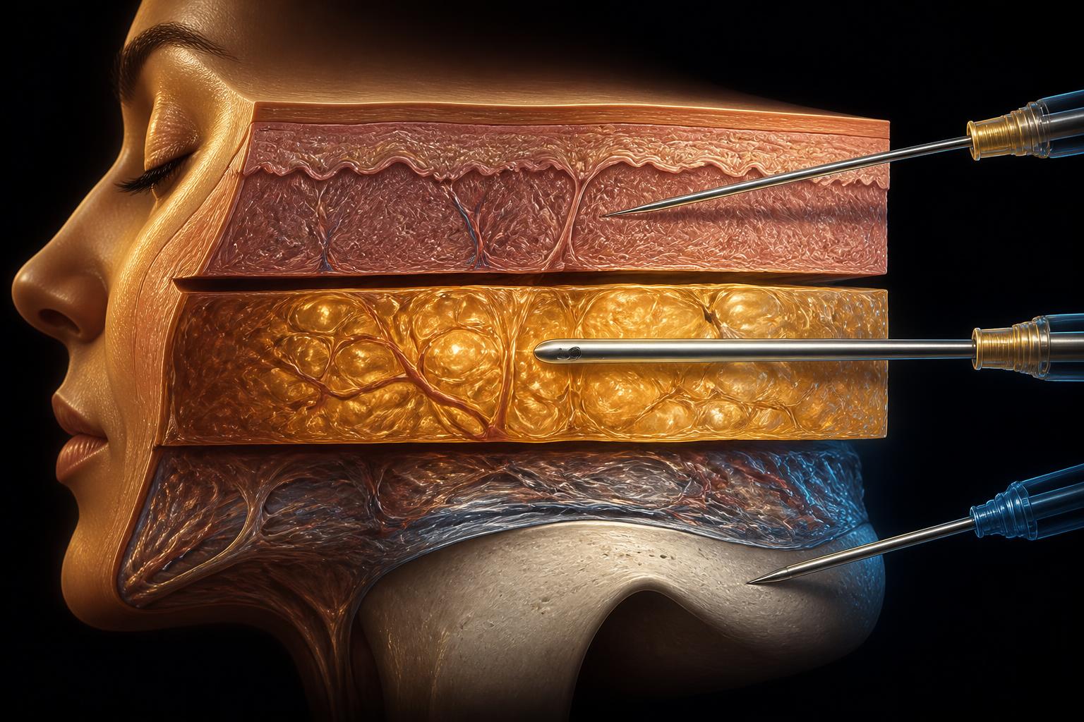
The injector cannot see the artery and cannot verify its path. What can be controlled is the plane the tip rests in — and that single variable organises both technique and safety.
Dr Ahmed Haq1
- 1 Founder and Medical Director, Harley Street Institute, London, United Kingdom
Corresponding author: journal@harleystreetinstitute.com
Journal: Aesthet Intell
DOI: to be assigned
Volume / Issue: 1 / 7
Pages: 115–128
Received: 2026-06-29
Accepted: 2026-07-06
Published: 2026-07-08
Licence: CC BY 4.0
Abstract
- Background.
- Every clinical discipline works with the anatomy its tools allow it to see. The surgeon confirms the course of a vessel under direct vision; the radiologist images the structure before anything is done. The aesthetic injector has neither — the needle works blind, and no amount of memorised anatomical variation can be verified at the moment of injection.
- Methods.
- Narrative review and educational synthesis of the published literature on facial vascular anatomy and variation, needle and cannula technique, injection depth, facial fat compartments and filler-related vascular complications, organised by treatment plane rather than by anatomical region.
- Results.
- What cannot be seen — the horizontal path of a vessel — must be surrendered; what can be controlled — the plane in which the tip rests — becomes the organising variable of safe practice. Filler practice resolves into three planes, each defined by its tool: the needle in the skin, the cannula in the subcutaneous plane, and the needle on the periosteum. The muscular plane is excluded as the territory of neuromodulators, and the deep fat compartments are excluded as a routine target on the grounds that anatomical ageing and treatment target are not the same thing.
- Conclusion.
- Organised around depth rather than path, filler practice becomes largely region-independent and more defensibly safe. The framework is offered as an educational and strategic scaffold, not as a substitute for supervised anatomical training.
Keywords: injection anatomy, dermal fillers, injection depth, cannula, facial rejuvenation, deep fat, aesthetic strategy
| Evidence domain | What the article reports | How to interpret it |
|---|---|---|
| Background | Every clinical discipline works with the anatomy its tools allow it to see. The surgeon confirms the course of a vessel under direct vision; the radiologist images the structure before anything is done. The aesthetic injector has neither — the needle works blind, and no amount of memorised anatomical variation can be verified at the moment of injection. | Defines the problem and the reason for review or investigation. |
| Methods | Narrative review and educational synthesis of the published literature on facial vascular anatomy and variation, needle and cannula technique, injection depth, facial fat compartments and filler-related vascular complications, organised by treatment plane rather than by anatomical region. | Shows how the evidence or observations were assembled. |
| Results | What cannot be seen — the horizontal path of a vessel — must be surrendered; what can be controlled — the plane in which the tip rests — becomes the organising variable of safe practice. Filler practice resolves into three planes, each defined by its tool: the needle in the skin, the cannula in the subcutaneous plane, and the needle on the periosteum. The muscular plane is excluded as the territory of neuromodulators, and the deep fat compartments are excluded as a routine target on the grounds that anatomical ageing and treatment target are not the same thing. | Summarises the principal findings reported by the authors. |
| Conclusion | Organised around depth rather than path, filler practice becomes largely region-independent and more defensibly safe. The framework is offered as an educational and strategic scaffold, not as a substitute for supervised anatomical training. | States the authors’ clinical interpretation and its limits. |
Postgraduate Level
Complete this article to earn your certificate
Read the article, complete a short assessment, and submit your reflection to receive your AiCE Points certificate. Equivalence to 1 CPD/CME point — we do not award CPD/CME directly.
Learning Objectives
- 1Explain why memorised vascular variation cannot be applied at the moment of injection
- 2Identify the three filler planes and the tool appropriate to each
- 3Justify the exclusion of the deep fat compartments as a routine treatment target
- 4Use plane selection, rather than vessel path prediction, as the primary vascular safety strategy
- 5Record injection depth as an anatomical plane paired with entry angle, not as a needle length
The Three-Layer Filler Model — Quick Reference
Layer 1 · Skin (dermis)
- Tool
- Needle.
- Intent
- Regenerate — hydration, skin boosters, fine surface lines.
- Product
- Soft, low volume.
- Risk
- Lumps and bluish discolouration if over-filled.
Layer 2 · Subcutaneous
- Tool
- Blunt cannula.
- Intent
- Contour — transitions, surface shape, soft-tissue support.
- Regions
- Cheek, jawline, tear trough, nasolabial.
- Why cannula
- Travels within the plane and displaces pliable vessels rather than piercing them.
Layer 3 · Periosteum
- Tool
- Needle to bone.
- Intent
- Project — skeletal-level structural support.
- Product
- Firm, cohesive bolus.
- Note
- Small deposits produce disproportionate surface change.
Excluded by design
- Muscle
- Territory of neuromodulators, not filler.
- Deep fat
- Not a routine target; treated selectively when its deficit dominates the deformity.
- Principle
- Anatomical ageing ≠ treatment target.
1. Introduction: every discipline has the anatomy its tools allow
Anatomy is not a single thing. It is shaped by what a practitioner is able to see and do. The surgeon is taught surgical anatomy: the field is opened, the tissues are separated under direct vision, and when a vessel does not follow its textbook course, the surgeon sees the variant and adapts to it in real time. The radiologist is taught radiological anatomy: the body is imaged first, and the intervention follows the picture. In both cases the anatomy that matters is the anatomy the practitioner can confirm before acting.[1]
The aesthetic injector has neither luxury. The needle passes beneath the skin unseen; there is no open field and, in routine practice, no live image. And here is the point that this paper turns on: even a complete knowledge of anatomical variation would not rescue the injector, because it cannot be applied at the moment it is needed. The facial artery, to take the most notorious example, follows a highly variable course; its path differs between individuals and between the two sides of the same face.[2,3] The injector cannot know, at the instant the needle enters, which variant lies beneath the chosen point of skin. Memorising every documented pathway of the facial artery therefore offers less protection than it appears to, because the one thing the injector needs — where is this particular vessel, in this particular patient, right now — is precisely the thing that cannot be seen.
What the injector can know, and control, is depth. The layer in which the tip comes to rest is not a matter of anatomical chance; it is a matter of technique, and it is reproducible. This is the central claim of the paper: for the injector, anatomy is depth. The discipline's curriculum, and its safety, should be organised around a single question asked at every injection — what layer am I in, and with what tool? — rather than around a horizontal map of named structures that cannot be verified through intact skin.
The model that follows is deliberately reductive, in the same spirit as the companion Three-Cell Model of skin rejuvenation.[4] It concerns fillers only. Botulinum toxin, which targets the muscular layer and acts on the neuromuscular junction, is a different treatment with a different logic and is not part of this model.[5]
2. The three layers of filler
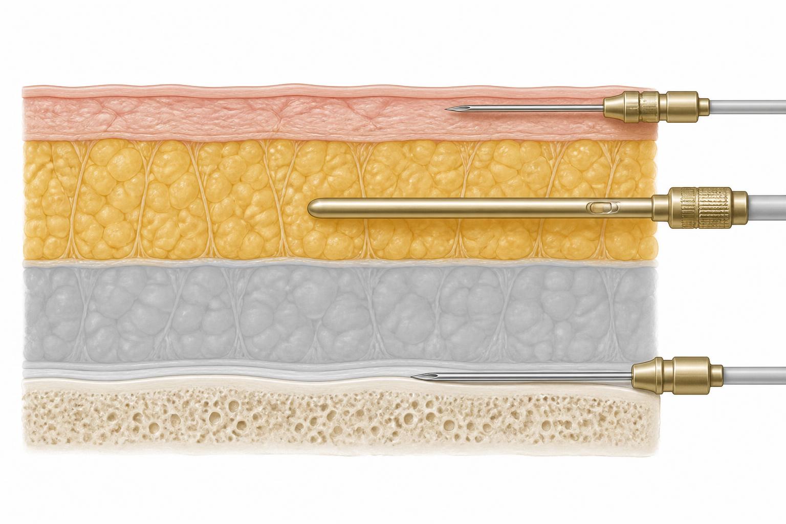
If the muscular plane is set aside as the territory of neuromodulators, the filler injector works, in practice, in three planes — and each is defined not only by its depth but by the tool appropriate to it. This pairing of layer and tool is the model.
Layer one — the needle, in the skin. The most superficial plane: the epidermis and, principally, the dermis. Here the work is fine and the volumes are minute — superficial hydration, skin boosters, the softening of fine surface lines. The tool is a needle, because the target is precise and shallow and a cannula cannot address it. Product must be soft and placed sparingly; the thin overlying tissue is unforgiving of any bulk, which shows through as a lump or a bluish discolouration.[6]
Layer two — the cannula, in the subcutaneous plane. Beneath the skin lies the subcutaneous space, and this is the injector's principal working layer for contour and soft-tissue support. The tool here is the blunt cannula, which travels within this plane over distance, pushing pliable structures — including vessels — aside rather than piercing them, and allowing a region to be treated through a single entry point.[7,8] This is the layer of transitions and surface shape: the cheek, the jawline, the tear trough and the nasolabial region are all, in this model, subcutaneous cannula work.
Layer three — the needle, on the periosteum. The deepest plane: the needle passed down to bone, depositing a firm bolus directly on the periosteum to provide structural, skeletal-level projection.[9] The tool is a needle, because the target is a precise point on bone and the product is firm and cohesive, chosen to resist the weight of the tissue above and to lift it. This is the plane of support and projection, not of contour.
The elegance of the model, and the reason it is worth teaching, is that it is largely independent of facial region. A filler behaves in these three planes according to the plane, not the location. A cannula in the subcutaneous plane of the nose is conceptually the same manoeuvre as a cannula in the subcutaneous plane of the cheek or the chin; a needle to periosteum at the zygoma follows the same logic as a needle to periosteum at the chin or the piriform aperture. The injector who has internalised the three layers does not need a separate mental protocol for every anatomical region. They need to decide, for the correction in front of them, which of the three planes it belongs in — and the tool, the product and the technique follow.
3. Why the deep fat is deliberately excluded
An anatomically minded reader will notice an omission. Between the subcutaneous plane and the periosteum lie the deep fat compartments — the deep medial cheek fat, the sub-orbicularis oculi fat, and others — which are the subject of a large and growing literature on midface ageing and volumisation.[10,11] Their exclusion from this model is deliberate, and it requires defence rather than silence.
The argument is not that the deep compartments do not age. They demonstrably do; longitudinal imaging shows measurable loss of deep midface fat with age, and in proportional terms that loss may exceed the loss in the superficial compartments.[12,13] The argument is a different and more important one: anatomical ageing and treatment target are not the same thing. That a structure has changed with age does not mean that replacing it, millilitre for millilitre, is the most efficient or the most elegant way to correct the visible result.
There is a sound biomechanical reason this holds. A small, well-placed deposit at the periosteal boundary alters the geometry of every layer stacked above it; the surface effect of a deep structural correction is disproportionate to its volume, and cadaveric surface-volume analyses confirm that compartments differ markedly in how much surface change a given volume produces.[14] In practice, this means that the aesthetic consequence commonly attributed to deep-fat deflation can frequently be corrected from the two ends of the stack — projection restored on the periosteum below, contour and transitions restored in the subcutaneous plane above — without entering the deep fat at all. Across nearly two decades of practice, I have found that treatment confined to the dermal, subcutaneous and periosteal planes is sufficient to produce a satisfying rejuvenation in the large majority of aesthetic patients, without routine recourse to deep-compartment volumisation.
This is best expressed as a hierarchy of intent. The model treats to regenerate the envelope (dermis), to contour the surface (subcutaneous), and to project the structure (periosteum) — regenerate, support, camouflage — rather than to identify each site of anatomical loss and replace it. Treatment volume need not equal age-related volume loss.
The caveat matters and is stated plainly. This is a default philosophy, not an absolute rule. There are phenotypes in which a deep-compartment deficit genuinely dominates the deformity — marked medial cheek deflation, certain infraorbital configurations, significant temporal hollowing — and in these, selective deep treatment is appropriate and sometimes necessary. Such cases sit at the more advanced or surgical end of practice, and they are the exception that the model accommodates rather than denies. Deep fat is not forbidden; it is simply not a primary or routine target, and it should be treated selectively, only when examination shows that its deficit is producing a visible problem that cannot be corrected more elegantly from the layers above and below it.
4. The safety argument: depth is more reliable than path
The layered model earns its keep most decisively in vascular safety, and here the paper's opening premise returns with full force. Intra-arterial injection of filler is the most consequential complication in the field; retrograde embolisation into the ophthalmic circulation can cause irreversible blindness, and occlusion of a cutaneous artery can cause tissue necrosis.[15,16] Preventing these events is the central safety task of injectable practice.[17]
The conventional response is to teach the injector the anatomical course of the dangerous vessels. This is necessary knowledge, but it is insufficient protection, for the reason established at the outset: the vessels vary, their paths differ between patients and between sides, and none of that variation can be seen through intact skin at the moment of injection.[2,3] An injector attempting to stay safe by predicting the exact path of the facial or angular artery is relying on a map that may not describe the patient in front of them.
Depth is the more reliable variable. While the horizontal path of a vessel varies, the plane in which the dangerous vessels of a region tend to run is considerably more consistent, and — crucially — the plane is something the injector controls rather than guesses.[18,19] The strategy that follows is therefore not “know where every artery runs,” which is unattainable in real time, but “know the safe plane for this region and remain deliberately within it.” In several regions the periosteal plane, deep beneath the level at which the named vessels travel, is comparatively safe for a bolus; in others the superficial subcutaneous plane, entered with a cannula, is preferred; the vessel-dense intermediate tissue is what the injector most often seeks to avoid.[9,18] The standard risk-reduction measures — injecting slowly and at low pressure, using small aliquots, preferring the cannula in broad high-risk planes, keeping the tip moving, and choosing the safe plane for the region — are all, at root, expressions of one principle: controlling depth. Path is what the injector cannot know; depth is what the injector can command.
5. Depth is a plane, not a needle length
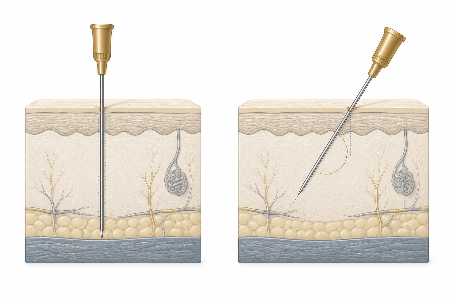
One clarification prevents a common error. To say a target lies “at five millimetres” is not to instruct the injector to bury five millimetres of needle. Deposition depth and needle-insertion depth are different quantities: the skin compresses on entry, so inserted length reaches deeper than the uncompressed measurement suggests; an oblique approach travels further through tissue to reach a given perpendicular depth; and tenting or pinching the tissue changes the relationship between surface and target.[20] The variable that governs both result and safety is the anatomical plane in which the tip finally rests — the layer — not the length of steel that has disappeared. The injector aims a tip at a layer, and it is the layer, not the ruler, that defines success.
6. Limitations
This is a teaching and strategic framework, with the limitations of any deliberate simplification. The planes of the face are not of uniform thickness across every region, and the model's region-independence is a teaching approximation rather than an anatomical identity; the transition between planes is subtler in some areas than others.[21] The relative safety of any plane is probabilistic, not absolute — no plane is reliably free of vessels, and catastrophic complications have occurred with careful technique in experienced hands.[16] The deep-fat position advanced here is a considered clinical philosophy supported by biomechanical reasoning and long personal experience; it would be strengthened by prospective, blinded, volumetric comparison of periosteal-plus-superficial correction against the same with added deep-compartment filler, which the author regards as a testable and worthwhile study. The framework is intended to organise the injector's reasoning and to be taught alongside — not instead of — supervised cadaveric and clinical anatomical training. It reduces risk; it does not remove it.
7. Conclusion
The injector cannot see the needle tip and cannot verify the anatomy beneath it, and so must reason about the one variable that is both knowable and controllable: depth. Organised around depth, filler practice resolves into three planes, each paired with its tool — the needle in the skin, the cannula in the subcutaneous, the needle on the periosteum — and this small model, largely indifferent to facial region, determines how the product behaves, which tool should deliver it, and how safely the injection is performed.
The model's most distinctive feature is what it leaves out. It does not chase the deep fat, because anatomical ageing is not the same as a treatment target: the face at sixty does not need to be reconstructed into the face at twenty, only to have the visible consequences of its ageing corrected — regenerated, supported and, where the skeleton has receded, strategically projected. That distinction, between reversing anatomy and managing its appearance, is the beginning of restraint, and restraint is the beginning of good aesthetic practice.
8. Conflict of interest
The author is the founder and Medical Director of the Harley Street Institute, which provides commercial aesthetic training, and is the author of a forthcoming book on aesthetic practice. This manuscript was handled independently of the journal's Editor-in-Chief.
9. Funding
None.
AI Disclosure
Drafting and reference formatting were assisted by an AI language model under the author's direction and review. The author is responsible for all clinical content and conclusions.
Competing Interests
The author(s) declare no competing financial or non-financial interests relevant to this work.
Funding
This work received no specific grant from any funding agency in the public, commercial, or not-for-profit sectors.
Ethics & Consent
Where applicable, ethical approval and informed patient consent were obtained in accordance with the Declaration of Helsinki. Reviews and commentaries did not require ethical approval.
HSI Editorial · Reflection & Forward Recommendations
Where we stand on this
Reflection
The injector's advantage is not a better map of the arteries. It is a better command of the plane.
Ask one question at every injection: what layer am I in, and with what tool?
Forward Recommendations
- Decide the plane before choosing the product, not after.
- Default to the cannula in broad subcutaneous work; reserve the needle for dermis and periosteum.
- Treat the deep fat only when examination shows its deficit is the dominant deformity.
- Record depth as a plane in your notes, never as a needle length.
Editorial position of the Harley Street Institute. Authored by the HSI Clinical Review Board; not a substitute for the peer-reviewed evidence summarised above.
The Anatomy of Injection
A three-part framework linking cellular biology, tissue planes and injection technique into a single teaching model.
- 1/3The Three-Cell Model of Skin Rejuvenation
- 2/3The Three-Layer Filler Model: Depth as the Anatomy of InjectionYou are here
- 3/3The HSI Mini Scale: Biological Sequencing of Skin, Movement, Support and Descent
References
- Standring S, editor. Gray's Anatomy: The Anatomical Basis of Clinical Practice. 42nd ed. London: Elsevier; 2020.
- Lohn JWG, Penn JW, Norton J, et al. The course and variation of the facial artery and vein. Surg Radiol Anat. 2011;33(10):841–850.
- Tansatit T, Apinuntrum P, Phetudom T. A typical pattern of the labial arteries with implications for lip augmentation with injectable fillers. Aesthetic Plast Surg. 2014;38(6):1083–1089.
- Haq A. The Three-Cell Model of Skin Rejuvenation: a simplified cellular framework for aesthetic practice. Aesthet Intell. 2026;1.
- de Almeida AT, Figueredo V, da Cunha ALG, et al. Consensus recommendations for the use of botulinum toxin. Dermatol Surg. 2017;43(Suppl 3):S217–S228.
- Micheels P, Sarazin D, Besse S, et al. A blanching technique for intradermal injection of hyaluronic acid. Plast Reconstr Surg. 2013;132(4 Suppl 2):59S–68S.
- Pavicic T, Frank K, Erlbacher K, et al. Precision in dermal filling: a comparison between needle and cannula. J Drugs Dermatol. 2017;16(9):866–872.
- Alam M, Kakar R, Dover JS, et al. Rates of vascular occlusion associated with using needles vs cannulas for filler injection. JAMA Dermatol. 2021;157(2):174–180.
- Surek CC, Beut J, Stephens R, et al. Pertinent anatomy and analysis for midface volumizing procedures. Plast Reconstr Surg. 2015;135(5):818e–829e.
- Rohrich RJ, Pessa JE. The fat compartments of the face: anatomy and clinical implications for cosmetic surgery. Plast Reconstr Surg. 2007;119(7):2219–2227.
- Gierloff M, Stohring C, Buder T, et al. Aging changes of the midfacial fat compartments: a computed tomographic study. Plast Reconstr Surg. 2012;129(1):263–273.
- Wysong A, Kim D, Joseph T, et al. Quantifying soft tissue loss in facial aging: a study using magnetic resonance imaging. Dermatol Surg. 2013;39(12):1895–1902.
- Zhang Y, Zhang J, Wang Y, et al. Facial aging: a quantitative analysis of midface volume changes over 11 years. Plast Reconstr Surg. 2021;147(1):11e–20e.
- Cotofana S, Gotkin RH, Frank K, et al. The surface-volume coefficient of the superficial and deep facial fat compartments: a cadaveric three-dimensional volumetric analysis. Plast Reconstr Surg. 2019;143(6):1605–1613.
- Beleznay K, Carruthers JDA, Humphrey S, et al. Update on avoiding and treating blindness from fillers: a recent review of the world literature. Aesthet Surg J. 2019;39(6):662–674.
- Goodman GJ, Roberts S, Callan P. Experience and management of intravascular injection with facial fillers. Aesthetic Plast Surg. 2016;40(4):549–555.
- DeLorenzi C. Complications of injectable fillers, part 2: vascular complications. Aesthet Surg J. 2014;34(4):584–600.
- Cotofana S, Lachman N. Arteries of the face and their relevance for minimally invasive facial procedures: an anatomical review. Plast Reconstr Surg. 2019;143(2):416–426.
- Scheuer JF, Sieber DA, Pezeshk RA, et al. Anatomy of the facial danger zones: maximizing safety during soft-tissue filler injections. Plast Reconstr Surg. 2017;139(1):50e–58e.
- Cotofana S, Alfertshofer M, Frank K, et al. Relationship between vertical anatomical planes and injection depth. Facial Plast Surg Aesthet Med. 2022;24(S1):S1–S8.
- Sandulescu T, Spilker L, Rauscher D, et al. The layered anatomy of the face revisited. Ann Anat. 2019;222:80–89.
Declarations
- Peer review:
- This article underwent single-blind external peer review by at least two independent reviewers, followed by editorial acceptance.
- Conflicts of interest:
- The author(s) declare no competing financial or commercial interests relating to the content of this article. Editorial decisions are made independently of the Harley Street Institute's commercial training activities.
- Funding:
- No external funding was received for the preparation of this article.
- Licence:
- © 2026 Harley Street Institute. Open access article distributed under the Creative Commons Attribution 4.0 International Licence (CC BY 4.0), permitting unrestricted use with appropriate citation.
Post-publication review
Discuss this article with the journal AI
Ask a clinical question about this article, or flag a possible error. Our AI agent will reply in real time, log your input, and — if you have identified a credible mistake — escalate it to the HSI editorial team for review and a published correction notice.
AI responses are generated by an assistant model. They do not constitute medical advice. Editorial corrections are only applied after a named HSI editor reviews and signs off.
Hi. I'm the Aesthetic Intelligence reader AI for this article.
Try: "Explain section 3 in plain English." · "What's the evidence for the 1-in-6,410 figure?" · "How does this compare to UK NICE guidance?"