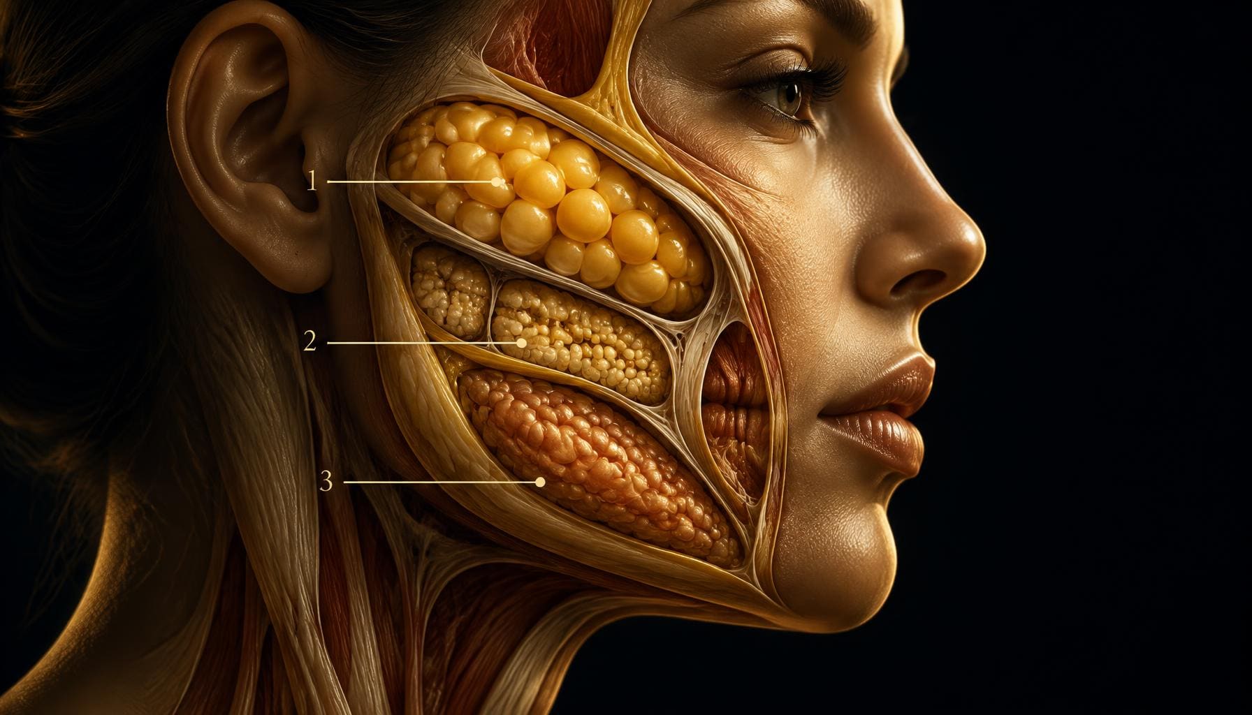
Review Article
Superficial Versus Deep Midfacial Fat: What Changes When the Buccal Fat Pad Is Counted Separately — and What Should That Mean for Filler Treatment?
Harley Street Institute · London
Aesthetic Intelligence · Peer-reviewed open access
Dr Ahmed Haq1
- 1 Founder and Medical Director, Harley Street Institute, London, United Kingdom
Corresponding author: journal@harleystreetinstitute.com
Journal: Aesthet Intell
DOI: to be assigned
Volume / Issue: 1 / 8
Pages: 207–222
Received: 2026-08-27
Accepted: 2026-08-29
Published: 2026-08-29
Licence: CC BY 4.0
Abstract
- Background.
- The division of facial adipose tissue into superficial and deep compartments has profoundly influenced modern injectable practice. A common therapeutic interpretation is that ageing preferentially reduces deep facial fat, causing secondary superficial descent and therefore favouring deep-first volumetric restoration. However, published estimates of the amount of “deep fat” vary considerably depending on which structures are included. In particular, grouping the relatively large buccal fat pad with the injectable-relevant deep midface compartments substantially changes the apparent superficial-to-deep ratio.
- Methods.
- This narrative review examines anatomical dissection and computed-tomographic studies quantifying superficial, deep and buccal facial fat. Particular attention is given to Raskin and LaTrenta, Mertens et al., Boehm et al., Tower et al., and longitudinal studies of superficial midfacial fat. Results are interpreted according to two separate definitions: the complete anatomical adipose system below the superficial musculoaponeurotic system (SMAS), and the more limited deep midfacial compartments commonly discussed as potential targets in injectable rejuvenation.
- Results.
- Whole-face dissection has reported approximately 57% of facial fat superficial to the SMAS and 43% below it. However, this broad anatomical calculation does not correspond directly to the deep compartments commonly considered for filler treatment. In the longitudinal CT study of Boehm et al., superficial midface fat averaged 26.10 mL, specifically defined deep midface fat 11.01 mL, and buccal fat 9.36 mL. When buccal fat is excluded from the comparison, superficial fat represents approximately 70% and specifically defined deep fat approximately 30% — a ratio of approximately 2.4:1. Mertens et al. independently reported a deep-to-superficial midface fat mass ratio of 0.44 in subjects aged 75 years or younger, mathematically corresponding to approximately 31% deep and 69% superficial tissue. Nevertheless, their older group demonstrated preferential deep-fat reduction, and Boehm et al. similarly found a larger proportional longitudinal decline in specifically defined deep fat. In contrast, Tower et al. found increasing rather than decreasing deep-cheek volume with age.
- Conclusion.
- Whether superficial and deep facial fat appear approximately equal or markedly different depends heavily on anatomical definition. For injectable medicine, the buccal fat pad should probably be reported separately rather than allowed to dominate calculations of “deep injectable fat.” Current evidence suggests that the specifically defined deep midfacial compartments may constitute substantially less volume than the superficial system while potentially undergoing proportionately greater age-related change. Neither observation establishes where filler must be placed. Anatomical ageing describes what has changed; treatment strategy requires a separate judgement about which change is clinically relevant.
Keywords: facial fat compartments, superficial fat, deep medial cheek fat, buccal fat pad, dermal filler, cannula, midface, facial ageing, volume loss, SMAS
| Evidence domain | What the article reports | How to interpret it |
|---|---|---|
| Background | The division of facial adipose tissue into superficial and deep compartments has profoundly influenced modern injectable practice. A common therapeutic interpretation is that ageing preferentially reduces deep facial fat, causing secondary superficial descent and therefore favouring deep-first volumetric restoration. However, published estimates of the amount of “deep fat” vary considerably depending on which structures are included. In particular, grouping the relatively large buccal fat pad with the injectable-relevant deep midface compartments substantially changes the apparent superficial-to-deep ratio. | Defines the problem and the reason for review or investigation. |
| Methods | This narrative review examines anatomical dissection and computed-tomographic studies quantifying superficial, deep and buccal facial fat. Particular attention is given to Raskin and LaTrenta, Mertens et al., Boehm et al., Tower et al., and longitudinal studies of superficial midfacial fat. Results are interpreted according to two separate definitions: the complete anatomical adipose system below the superficial musculoaponeurotic system (SMAS), and the more limited deep midfacial compartments commonly discussed as potential targets in injectable rejuvenation. | Shows how the evidence or observations were assembled. |
| Results | Whole-face dissection has reported approximately 57% of facial fat superficial to the SMAS and 43% below it. However, this broad anatomical calculation does not correspond directly to the deep compartments commonly considered for filler treatment. In the longitudinal CT study of Boehm et al., superficial midface fat averaged 26.10 mL, specifically defined deep midface fat 11.01 mL, and buccal fat 9.36 mL. When buccal fat is excluded from the comparison, superficial fat represents approximately 70% and specifically defined deep fat approximately 30% — a ratio of approximately 2.4:1. Mertens et al. independently reported a deep-to-superficial midface fat mass ratio of 0.44 in subjects aged 75 years or younger, mathematically corresponding to approximately 31% deep and 69% superficial tissue. Nevertheless, their older group demonstrated preferential deep-fat reduction, and Boehm et al. similarly found a larger proportional longitudinal decline in specifically defined deep fat. In contrast, Tower et al. found increasing rather than decreasing deep-cheek volume with age. | Summarises the principal findings reported by the authors. |
| Conclusion | Whether superficial and deep facial fat appear approximately equal or markedly different depends heavily on anatomical definition. For injectable medicine, the buccal fat pad should probably be reported separately rather than allowed to dominate calculations of “deep injectable fat.” Current evidence suggests that the specifically defined deep midfacial compartments may constitute substantially less volume than the superficial system while potentially undergoing proportionately greater age-related change. Neither observation establishes where filler must be placed. Anatomical ageing describes what has changed; treatment strategy requires a separate judgement about which change is clinically relevant. | States the authors’ clinical interpretation and its limits. |
Postgraduate Level
Complete this article to earn your certificate
Read the article, complete a short assessment, and submit your reflection to receive your AiCE Points certificate. Equivalence to 1 CPD/CME point — we do not award CPD/CME directly.
Learning Objectives
- 1Distinguish superficial midfacial fat, specifically defined deep midfacial fat and the buccal fat pad
- 2Explain why grouping buccal fat with the deep compartments changes the apparent superficial-to-deep ratio
- 3Interpret the 57:43 and approximately 70:30 figures without treating either as a universal constant
- 4Separate absolute compartment volume from proportional age-related volume loss
- 5Weigh competing evidence for deep-fat atrophy, stability and apparent volume increase
- 6Decide independently whether a demonstrated anatomical change is a clinically appropriate filler target
HSI Quick Reference
The two ratios
- 57 : 43
- All fat above vs below the SMAS, buccal fat included — a broad anatomical figure, not an injectable one
- 70 : 30
- Superficial vs specifically defined deep midface fat when the buccal fat pad is reported separately (Boehm; Mertens ≈ 69:31)
- Ratio
- ≈ 2.4 : 1 superficial to deep, excluding buccal fat
Boehm baseline volumes
- Superficial midface fat
- 26.10 mL
- Specifically defined deep fat
- 11.01 mL
- Buccal fat pad
- 9.36 mL — best shown in its own column
Ageing
- Proportional loss
- Deep fat lost a greater percentage of its starting volume (18.4% vs 11.3% over 11.3 years)
- Absolute loss
- Superficial fat lost more volume in millilitres (2.95 vs 2.03 mL)
- Contradiction
- Tower et al. found deep-cheek volume increased with age — possibly buccal pseudoherniation
HSI principle
- Should anatomy determine filler allocation?
- No
- Core rule
- Anatomical ageing is not the same as a treatment target
Learning Objectives
After reading this review, the practitioner should be able to: distinguish superficial midfacial fat, specifically defined deep midfacial fat and the buccal fat pad; understand why grouping buccal fat with all other deep fat changes the apparent superficial-to-deep ratio; interpret the 57:43 and approximately 70:30 figures without treating either as a universal anatomical constant; distinguish absolute compartment volume from proportional age-related volume loss; compare competing evidence for deep-fat atrophy, stability, redistribution and apparent volume increase; and decide independently whether a demonstrated anatomical change represents a clinically appropriate filler target.
This article carries 1 AiCE Point at postgraduate level.
1. Clinical Context: One Word, Several Different Anatomies
“Deep fat” sounds like a single anatomical category. It is not.
At a simple five-layer level the face can be described as: skin; superficial/subcutaneous fat; SMAS and mimetic muscular layer; deep fat and deep spaces; and periosteum/deep fascia and skeleton. This is useful for teaching, but it can hide important distinctions inside layer four.
The deeper facial adipose system contains structures that differ substantially in location, function and relevance to injectable medicine. These include deep medial cheek fat, suborbicularis oculi fat (SOOF), other region-specific deep compartments and the buccal fat pad.
The buccal fat pad deserves particular attention because it is comparatively large. When it is automatically placed in the same numerical basket as the smaller deep compartments routinely discussed in filler treatment, the resulting number answers the anatomical question: how much facial adipose tissue lies beneath the SMAS? It does not necessarily answer the clinical question: how large are the deep compartments that an aesthetic injector might reasonably consider restoring? Those are different questions.
26.10 mL
Superficial midface fat
Boehm et al., baseline CT volume
11.01 mL
Deep midface fat
Specifically defined injectable compartments
9.36 mL
Buccal fat pad
Reported separately in this review
2.37:1
Superficial : deep
With buccal fat kept out of the comparison
2. Why the Buccal Fat Pad Should Be Shown Separately
The buccal fat pad is unquestionably a deep facial adipose structure. There is therefore nothing anatomically incorrect about including it in a description of the total adipose tissue beneath the SMAS. The problem occurs when the resulting number is subsequently interpreted as though all of this “deep volume” were comparable from an injectable perspective.
In the Boehm study, specifically defined deep midface fat averaged 11.01 mL, while buccal fat averaged 9.36 mL.[4] If these are combined, deep + buccal = 20.37 mL — and the buccal fat therefore contributes approximately 46% of the combined deeper volume. In other words, almost half of what becomes labelled “deep fat” in the combined calculation is the buccal fat pad. Yet this is not ordinarily what an injector means when discussing routine restoration of deep medial cheek fat or SOOF.
For that reason, this review proposes reporting the buccal fat pad in a third column, rather than pretending it does not exist or automatically combining it with everything deep to the SMAS.
- Deep midface compartments (11.01 mL)54%
- Buccal fat pad (9.36 mL)46%
Source: Boehm et al., baseline volumes as reported in the text.
3. Two Correct Answers to Two Different Questions
This framing allows the apparently contradictory numbers to coexist.
Question 1: how much total facial adipose tissue is superficial versus beneath the SMAS? Raskin and LaTrenta performed anatomical dissections of ten fresh hemicadavers and reported approximately 57% above the SMAS and 43% below it.[1] That is a useful broad anatomical observation. It tells us that deep facial adipose tissue as a whole is not trivial. It does not tell us that 43% of an aesthetic treatment should be deep.
Question 2: how much of the measured midface consists of superficial fat compared with specifically defined deep midface fat, if buccal fat is kept separate? Using Boehm's baseline measurements — superficial 26.10 mL, deep 11.01 mL — the comparison becomes 70.3% superficial versus 29.7% deep, a superficial-to-deep ratio of approximately 2.37:1.[4] That is arguably the more relevant number when discussing the relative volumes of the two principal fat systems commonly considered during midfacial injectable planning.
Neither figure invalidates the other. They simply describe different denominators.
Buccal fat counted as deep
- Superficial (26.10 mL)56%
- Deep + buccal (20.37 mL)44%
Buccal fat reported separately
- Superficial (26.10 mL)70%
- Deep midface (11.01 mL)30%
Source: Boehm et al. (volumes); Raskin & LaTrenta (whole-face 57:43 dissection figure).
Source: Boehm et al., baseline measurements.
4. A Second Study Produces a Surprisingly Similar Relationship
Mertens, Foyatier and Mojallal dissected superficial and deep midfacial fat compartments in 40 cadavers. The investigators specifically described the deep midfacial system in terms including deep medial cheek fat and SOOF rather than simply treating every piece of fat under the SMAS as an equivalent unit.[2]
In subjects aged 75 years or younger they reported a deep-to-superficial mass ratio of 0.44. A ratio is not itself a percentage, but converting it mathematically: deep fraction = 0.44 ÷ 1.44 = 30.6%; superficial fraction = 1 ÷ 1.44 = 69.4% — approximately 69% superficial to 31% deep. That is remarkably close to the 70:30 relationship derived from Boehm when buccal fat is kept separate.
This convergence is interesting. It does not establish a universal physiological law that the midface is always exactly 70% superficial and 30% deep — the studies used different methodologies; one measured cadaveric mass, one measured CT-derived volume; populations, anatomical boundaries, age and BMI differed. But it suggests that approximately two-thirds to three-quarters superficial versus one-quarter to one-third specifically defined deep midfacial fat is a reasonable hypothesis for further investigation. That is considerably different from the mental image produced by 57:43.
Two independent datasets, one similar relationship
A deep-to-superficial ratio is converted to percentages as deep = r ÷ (1 + r).
| Study | Method | Reported figure | Superficial : deep |
|---|---|---|---|
| Raskin & LaTrenta | Dissection, 10 fresh hemicadavers, whole face | 57% above SMAS / 43% below | 57 : 43 (all sub-SMAS fat) |
| Boehm et al. | Longitudinal CT, midface volumes | 26.10 mL vs 11.01 mL | 70 : 30 (buccal excluded) |
| Mertens et al. (≤75 years) | Cadaveric dissection, 40 subjects, mass | Deep : superficial ratio 0.44 | 69 : 31 |
| Mertens et al. (>75 years) | Cadaveric dissection, 40 subjects, mass | Deep : superficial ratio 0.20 | 83 : 17 |
The figures are not contradictory; they answer different anatomical questions with different denominators.
5. Age Changes the Ratio Again
Mertens' older group makes the picture even more interesting. Among subjects older than 75 years, mean deep-compartment mass was 1.48 g compared with 3.41 g in the younger group. The superficial compartments did not show a statistically significant corresponding decline. The deep-to-superficial ratio fell from 0.44 to 0.20[2] — a ratio of 0.20 corresponding mathematically to approximately 83% superficial and 17% deep within the compartments they measured.
This was why the authors concluded that deep midfacial fat undergoes preferential age-related atrophy and argued that restoring deep volume should be considered a priority. That interpretation deserves to be presented fairly: the data do provide an anatomical argument for selective deep-fat loss. But another conclusion is equally important — the compartment that loses the greater percentage is not necessarily the compartment containing the greater absolute volume. These are different concepts.
Source: Mertens, Foyatier & Mojallal, as reported in the text.
6. Absolute Volume Versus Percentage Loss
This distinction is easy to miss. Consider the Boehm longitudinal data over an average 11.3 years. Superficial fat fell from 26.10 to 23.15 mL — a loss of 2.95 mL, or 11.3%. Deep fat fell from 11.01 to 8.98 mL — a loss of 2.03 mL, or 18.4%.[4]
Therefore deep fat lost more as a percentage of its starting size, but superficial fat lost more absolute volume. Both statements are true. Which one matters more clinically cannot be answered from the CT numbers alone. If deep fat provides mechanically important support, a smaller absolute change could have an outsized surface effect. Alternatively, because superficial fat lies immediately beneath the visible surface, a change in superficial contour may be aesthetically important despite a smaller proportional loss. The imaging study does not determine which interpretation should govern filler treatment. That requires clinical outcome research.
If buccal fat remains separate, Boehm's follow-up measurements were superficial 23.15 mL, deep 8.98 mL and buccal 8.68 mL.[4] Considering only superficial versus specifically defined deep fat: 72.1% superficial versus 27.9% deep. The superficial-to-deep ratio therefore changes from approximately 2.37:1 to 2.58:1 over the observation period — the deep compartment became proportionately smaller.
Again, there are two possible ways to read this. One practitioner may conclude that deep fat is relatively small and preferentially atrophies, and therefore that replacing a limited amount of deep volume may efficiently restore support. Another may conclude that, despite preferential deep loss, the superficial system remains by far the larger adipose layer, so one should not assume every visible contour defect requires deep reconstruction. The evidence permits both hypotheses. What it does not permit is claiming that either treatment philosophy has been proven superior.
7. A Contradictory Longitudinal Study
This becomes more important when Tower, Gordon and Paskhover are considered. Their CT study included 109 patients cross-sectionally, with 23 subjects also having longitudinal imaging over an average 10.3 years. Rather than finding deep-cheek loss, they found an average 0.23-mL increase in deep cheek volume; increasing age was also associated with increasing deep cheek volume in the cross-sectional analysis.[3]
The authors proposed that this may reflect pseudoherniation or migration of buccal fat rather than true adipose generation. That possibility is itself revealing. If neighbouring adipose tissue can migrate or protrude into a radiologically defined deep-cheek region, then measured compartment volume represents not only atrophy but potentially position, boundaries, redistribution and tissue movement. This complicates any model based solely on “fat loss.”
8. Superficial Fat Also Refuses to Behave Simply
Superficial facial fat is equally heterogeneous. One longitudinal study following 14 patients over approximately ten years found no statistically significant change in overall superficial cheek-fat volume. A larger cross-sectional CT study of 109 patients found mean superficial cheek volume of 10.46 cc, with age, sex and BMI together accounting for approximately half of the variance; BMI was an especially important predictor.[5]
Therefore the superficial compartment should not simply be described as “the fat that falls because the deep fat disappears.” Age-related superficial morphology may involve local atrophy, relative preservation, inferior redistribution, enlargement of selected lower compartments, body-weight effects, sex differences, ligamentous restriction and gravitational displacement. The individual patient may show more than one of these processes simultaneously.
9. The More Useful Model: Three Adipose Categories
For injectable education we therefore favour depicting the midface as three adipose categories, not merely superficial versus deep. First, superficial fat — the superficial medial cheek, middle cheek, nasolabial and infraorbital/lateral superficial compartments — frequently encountered in contouring and blending. Second, specifically defined deep midface fat — deep medial cheek fat and selected SOOF/deep compartments — a potential selective treatment target. Third, the buccal fat pad and its extensions — important anatomy, but generally not a routine hyaluronic-acid augmentation target.
This prevents a very large buccal structure from silently changing the answer to a question about injectable deep-fat treatment. It also avoids the opposite mistake: pretending buccal fat is anatomically irrelevant.
Excluding the buccal pad from an injectable-volume comparison does not mean excluding it from facial ageing. The buccal fat pad can influence cheek fullness, lower-midface contour, the relationship between anterior and lateral facial volume, deep-space anatomy, and apparent changes caused by displacement or pseudoherniation. Tower's suggestion that apparent deep-cheek volume increase could reflect buccal-fat pseudoherniation is an excellent example.[3] The correct formulation is: separate, not ignore. That is the key distinction.
10. Does 70:30 Support Superficial Cannula Treatment?
This is where restraint is important. The 70:30 observation is compatible with a superficial-treatment philosophy. It does not validate one.
The superficial system is larger in the studies discussed above and lies immediately beneath the visible surface. There is therefore a plausible anatomical explanation for why small amounts of superficial correction can produce substantial changes in contour. But the deep system may have a different mechanical role per millilitre — a smaller deep compartment could conceivably provide more structural influence than its absolute volume suggests. Volume is not the same as mechanical importance. This is one of the most important ideas in this review.
In the author's practice, broad facial filler contouring is performed predominantly within a superficial subcutaneous cannula plane, with deep-fat or periosteal treatment used selectively when an identifiable structural indication exists. This differs from a routine “deep first” philosophy. The clinical observation behind the approach is simple: in many patients, correction of the visible contour immediately beneath the surface appears sufficient without attempting to reproduce the presumed volume of every deeper anatomical compartment. This is an author technique and clinical hypothesis, not a conclusion proven by the volume studies reviewed here — and that distinction should remain explicit. The anatomy provides plausibility. It does not provide comparative efficacy evidence.
11. Mid-Cheek: What Are We Actually Correcting?
The mid-cheek illustrates why this matters. A visible groove can result from several different mechanisms: superficial compartment contour difference; deep-fat reduction; skeletal projection change; retaining-ligament tethering; relative tissue descent; neighbouring tissue expansion; previous filler; or combinations of the above.
The temptation is to see a depression and label it “lost volume.” But a depression describes a surface geometry; it does not identify its anatomical cause. The clinician should therefore ask what changes when the tissue is examined, moved, animated and viewed from different angles. If a small superficial correction resolves the transition naturally, one may question what additional benefit would be gained by deeper replacement. If it does not, deeper support may be important. The sequence should emerge from the phenotype rather than from an anatomical doctrine.
12. Tear Trough: An Even Better Example
The medial tear trough illustrates the danger of equating anatomical terminology with a treatment target. SOOF is frequently mentioned in under-eye treatment, but the true medial tear trough does not simply sit above a large, uniform SOOF compartment; the deeper suborbicularis fat system becomes more anatomically relevant laterally. Therefore a superficial medial tear-trough correction should not automatically be described as “replacing SOOF.” A clinician may instead be altering the visible transition close beneath the skin — and the result may be successful without representing anatomical restoration.
This gives us another useful distinction: correction does not prove replacement. If an aesthetic defect improves after modifying one layer, that does not demonstrate that the ageing process originated within that same layer. Conversely, showing that ageing occurred in another layer does not prove that it had to be directly replaced.
13. What About the “Deep First” Philosophy?
The deep-first philosophy has a coherent anatomical basis. Mertens found preferential reduction of deep-fat mass in older cadavers and explicitly concluded that deep restoration should be considered a priority.[2] Boehm showed proportionally greater deep than superficial loss and interpreted this as support for the pseudoptosis hypothesis.[4] It would be inappropriate to dismiss these findings.
But the evidence does not yet answer the next therapeutic question: does treating the deep compartment first result in a better aesthetic outcome than treating an appropriate superficial target first? Those are not anatomical experiments. They are treatment-comparison experiments. And they largely remain to be performed.
14. What the Evidence Shows — and What It Does Not
The current literature allows several reasonably strong observations. First, superficial midfacial fat constitutes a substantial — probably predominant — proportion of the specifically measured superficial-plus-deep midface compartments when the buccal fat pad is kept separate. Second, selected deep midface compartments appear to undergo significant age-related change and may lose a greater percentage of their initial volume in some studies. Third, absolute superficial volume change may nevertheless equal or exceed absolute deep volume change. Fourth, the buccal fat pad can materially distort a simplistic superficial-versus-deep calculation. Fifth, other longitudinal research has observed increasing rather than decreasing deep-cheek volume, demonstrating that distribution and compartment boundaries complicate a pure-deflation model. Sixth, BMI, sex and individual anatomy substantially affect superficial volume. None of these observations establishes an obligatory filler sequence.
Equally, the literature does not currently prove that every ageing face loses deep fat; that every mid-cheek groove is caused by deep-fat loss; that the percentage of native tissue should determine filler allocation; that a 30% deep-fat fraction means 30% of injected product belongs deep; that deep replacement should universally precede superficial contouring; that superficial correction is universally superior; that successful treatment of a surface contour demonstrates that the pathology was superficial; or that anatomical restoration is synonymous with aesthetic improvement. That is where practitioner reasoning must begin.
15. Anatomy Is a Map, Not a Prescription
The most useful way to teach the numbers may therefore be as follows. Anatomical system: if all fat below the SMAS is counted, including the buccal fat pad, approximately 57:43 superficial to deep has been reported.[1] Injectable midface comparison: if the buccal pad is reported separately, approximately 70:30 superficial to specifically defined deep midface fat appears in two different datasets.[2,4] Ageing: some studies suggest deep compartments undergo greater proportional atrophy — but other studies show deep-cheek volume can remain stable or increase.[3] Treatment: therefore no anatomical ratio determines an injection ratio.
That is probably the most honest way to present the literature.
16. Five Questions Before Adding Volume
Rather than instructing the practitioner to inject one plane first, the HSI framework asks five questions.
First: what exactly is the visible phenotype — hollow, groove, flattening, transition, heaviness, descent or asymmetry? Second: which layer is most likely producing it — skin, superficial fat, muscle, deep fat, skeleton, ligamentous fixation, or several? Third: is tissue actually missing, or has it changed position? Fourth: what is the smallest intervention likely to change the visible phenotype — the treatment target need not reproduce the anatomy that existed at age twenty? Fifth: what remains after reassessment — another plane should be added because a residual problem remains, not because a theoretical sequence says it comes next.
A related principle deserves emphasis because it is easily forgotten: native volume is not treatment volume. A native fat compartment consists of living adipocytes, fibrous septae, vascular structures, extracellular matrix and tissue relationships that differ fundamentally from an implanted hyaluronic-acid gel. Therefore 1 mL of lost native fat does not equal 1 mL of required HA filler — neither shape nor mechanics are equivalent. A radiological measurement of volume change is useful for understanding ageing but cannot be converted directly into a syringe calculation. Even a perfect study proving that a compartment lost 2 mL would not establish that the patient should receive 2 mL of filler there.
17. The Study That Would Actually Answer the Clinical Question
The next important research question is not simply another cadaver dissection. It is a comparative treatment study. Patients with a reasonably homogeneous mid-cheek phenotype could be prospectively assigned to strategies such as predominantly superficial contour correction, selectively deep structural correction, or combined deep and superficial treatment.
The important endpoints would include blinded aesthetic assessment, three-dimensional surface change, FACE-Q or equivalent patient-reported outcome, total filler volume, ultrasound-confirmed product location, movement and expression, oedema, durability, naturalness, incidence of visible or palpable product, and evidence of overcorrection.
The most interesting question would be: which strategy produces the required visible change with the least total intervention and least alteration of facial identity? Until such comparative work exists, anatomy should guide judgement rather than replace it.
18. Conclusion
How much facial fat is superficial and how much is deep? The most accurate answer is: first define “deep.”
When every adipose structure below the SMAS is placed into one category, including the buccal fat pad, superficial and deeper tissue may appear relatively similar in overall quantity. When the buccal fat pad is separated — as Boehm and colleagues themselves did — the picture changes substantially. In their midface CT data, superficial fat measured 26.10 mL compared with 11.01 mL of specifically defined deep fat: approximately 70% superficial versus 30% deep. Mertens' independent anatomical study produces a surprisingly similar relationship in younger and middle-aged cadavers, although it also provides important evidence for preferential deep-fat reduction in advanced age.
The ageing story therefore cannot be reduced to one ratio. Deep fat may be smaller in absolute volume yet lose a greater proportion of itself. Superficial fat may remain the larger system while simultaneously redistributing. Buccal fat may alter apparent deep-cheek volume without being an ordinary injectable target. BMI, sex and individual anatomy modify all of these relationships.
The correct lesson is not “fill superficial fat because there is more of it,” nor “fill deep fat because it loses proportionally faster.” The useful conclusion is: understand which tissues have changed, establish which change is producing the patient's visible phenotype, and then decide whether that anatomical change is actually worth treating. Anatomy tells us what exists. Ageing research tells us what may change. Neither, by itself, tells us where the syringe should go. That final step remains clinical judgement. Anatomical ageing is not the same as a treatment target.
For the practical anatomy that follows from this analysis — superficial subcutaneous compartments, retaining ligaments, deep fat, tear-trough anatomy, cannula behaviour and the Three-Layer Filler Model — readers are directed to the Aesthetic Talk series, where the procedural detail is developed in full.
AI Disclosure
AI-assisted tools were used to organise the comparative calculations and cross-check ratio conversions against the published source data. All anatomical interpretation, clinical reasoning and conclusions are the author's own and have undergone editorial review.
Competing Interests
The author(s) declare no competing financial or non-financial interests relevant to this work.
Funding
This work received no specific grant from any funding agency in the public, commercial, or not-for-profit sectors.
Ethics & Consent
Where applicable, ethical approval and informed patient consent were obtained in accordance with the Declaration of Helsinki. Reviews and commentaries did not require ethical approval.
HSI Editorial · Reflection & Forward Recommendations
Where we stand on this
Reflection
The fat-compartment literature has given aesthetic medicine a much more sophisticated map of the face. The unintended consequence may have been a tendency to turn every labelled structure into an injectable target.
Perhaps the next stage in aesthetic anatomy is not discovering another compartment. It is learning when not to replace one.
Forward Recommendations
- Report the buccal fat pad separately whenever superficial and deep midfacial fat are compared — it contributes almost half of the combined “deep” volume and is not a routine augmentation target.
- Distinguish absolute volume change from proportional volume change before drawing any treatment conclusion from imaging data.
- Name the layer producing the visible phenotype before selecting a plane; a depression describes a surface geometry, not an anatomical cause.
- Never convert a radiological volume deficit directly into a syringe calculation — native tissue and implanted gel are not mechanically equivalent.
- Treat deep-first and superficial-first philosophies as hypotheses awaiting comparative outcome studies, not as settled doctrine.
Editorial position of the Harley Street Institute. Authored by the HSI Clinical Review Board; not a substitute for the peer-reviewed evidence summarised above.
References
- Raskin E, LaTrenta GS. Why do we age in our cheeks? Aesthet Surg J. 2007;27(1):19-28.
- Mertens A, Foyatier JL, Mojallal A. Quantitative analysis of midface fat compartments mass with ageing and body mass index, anatomical study. Ann Chir Plast Esthet. 2016;61(6):798-805.
- Tower JI, Gordon NA, Paskhover B. Deep cheek fat volumes and midfacial aging. Aesthet Surg J. 2020;40(5):467-475.
- Boehm LM, Morgan A, Hettinger P, Matloub HS. Facial aging: a quantitative analysis of midface volume changes over 11 years. Plast Reconstr Surg. 2021;147(2):319-327.
- Tower JI, Gordon NA, Paskhover B. Patterns of superficial midfacial fat volume distribution differ by age and body mass index. Aesthetic Plast Surg. 2019;43(5):1181-1189.
Declarations
- Peer review:
- This article underwent single-blind external peer review by at least two independent reviewers, followed by editorial acceptance.
- Conflicts of interest:
- The author(s) declare no competing financial or commercial interests relating to the content of this article. Editorial decisions are made independently of the Harley Street Institute's commercial training activities.
- Funding:
- No external funding was received for the preparation of this article.
- Licence:
- © 2026 Harley Street Institute. Open access article distributed under the Creative Commons Attribution 4.0 International Licence (CC BY 4.0), permitting unrestricted use with appropriate citation.
Post-publication review
Discuss this article with the journal AI
Ask a clinical question about this article, or flag a possible error. Our AI agent will reply in real time, log your input, and — if you have identified a credible mistake — escalate it to the HSI editorial team for review and a published correction notice.
AI responses are generated by an assistant model. They do not constitute medical advice. Editorial corrections are only applied after a named HSI editor reviews and signs off.
Hi. I'm the Aesthetic Intelligence reader AI for this article.
Try: "Explain section 3 in plain English." · "What's the evidence for the 1-in-6,410 figure?" · "How does this compare to UK NICE guidance?"