
Review Article · Aesthetic Intelligence · Vol 1 · Issue 4
Vascular Complications in Aesthetic MedicineThe First Hour Is the Whole Game
Vascular occlusion is rare, recognisable and — with a written, rehearsed protocol executed in the first hour — survivable without permanent harm. Readiness is a property of the clinic, not the injector.
HSI Team1
- 1 Harley Street Institute, London, United Kingdom
Corresponding author: journal@harleystreetinstitute.com
Journal: Aesthet Intell
DOI: to be assigned
Volume / Issue: 1 / 4
Pages: 52–72
Received: 2025-09-04
Accepted: 2025-10-22
Published: 2026-05-21
Licence: CC BY 4.0
From the Editor's Desk
A vascular occlusion does not give you the courtesy of a warning. It does not announce itself. It does not wait until the patient has paid and left. It happens, with a particular small wet hiss of dread, while you still have the syringe in your hand, and the rest of your career is decided in the next hour by what is already written down — or not — in the desk drawer beside you.
The clinics that lose patients to this are not the clinics with the worst injectors. They are the clinics with no protocol, no rehearsed sequence, no hyaluronidase on the shelf, and no named ophthalmologist on speed-dial. Skill matters. Preparation matters more. The next ten thousand words are about turning the worst hour of your professional life into a piece of paper you have already read.
Abstract
- Background.
- Intra-arterial injection of dermal filler is the single most consequential complication in non-surgical aesthetic medicine. Although rare per millilitre injected, it carries the potential for skin necrosis, scarring and — in the case of retrograde embolisation to the ophthalmic circulation — irreversible blindness (Beleznay et al., 2019; Goodman et al., 2020). The window between event and irreversible tissue loss is short, and outcome is determined almost entirely by whether a written, rehearsed emergency protocol is executed within hours, not days.
- Methods.
- Narrative review of the published peer-reviewed literature on vascular adverse events with hyaluronic acid (HA) and non-HA fillers (2014–2025), including the DeLorenzi high-dose pulsed hyaluronidase protocol, the ASDS Multi-Society guideline (Alam et al., 2021), the Beleznay vision-loss case series, and IMCAS / Aesthetic Complications Expert (ACE) Group consensus statements. The review is supplemented by a fully consented clinical case from an HSI Team member illustrating the natural history of a delayed-presentation lip and naso-labial vascular occlusion over 0, 24 hours, 72 hours and 3 months.
- Results.
- A three-stage operational framework — Prevent, Recognise, Treat — supports immediate clinical decision-making. Prevention rests on anatomical knowledge by plane, slow low-pressure delivery, small aliquots, aspiration where mechanically meaningful and cannula use in high-risk zones. Recognition is dominated by disproportionate pain, immediate blanching, dusky reticular livedo, delayed capillary refill and, most urgently, any visual symptom. Treatment is high-dose pulsed hyaluronidase delivered into and around the ischaemic territory, repeated hourly until reperfusion, supported by warm compresses, topical 2% glyceryl trinitrate (where evidence is contested), aspirin, oral steroids, broad-spectrum antibiotic cover for compromised skin, and immediate referral to hyperbaric oxygen and ophthalmology where indicated.
- Conclusion.
- Every aesthetic clinic should hold a written, rehearsed, in-date vascular emergency protocol; stock at least 3000 units of hyaluronidase on-site; maintain a named ophthalmology and hyperbaric pathway; and treat the first hour as the operative window. Outcome correlates with preparedness, not seniority.
Keywords: vascular occlusion, hyaluronidase, dermal filler complications, retrograde embolisation, superior labial artery, angular artery, hyperbaric oxygen, patient safety
| Evidence domain | What the article reports | How to interpret it |
|---|---|---|
| Background | Intra-arterial injection of dermal filler is the single most consequential complication in non-surgical aesthetic medicine. Although rare per millilitre injected, it carries the potential for skin necrosis, scarring and — in the case of retrograde embolisation to the ophthalmic circulation — irreversible blindness (Beleznay et al., 2019; Goodman et al., 2020). The window between event and irreversible tissue loss is short, and outcome is determined almost entirely by whether a written, rehearsed emergency protocol is executed within hours, not days. | Defines the problem and the reason for review or investigation. |
| Methods | Narrative review of the published peer-reviewed literature on vascular adverse events with hyaluronic acid (HA) and non-HA fillers (2014–2025), including the DeLorenzi high-dose pulsed hyaluronidase protocol, the ASDS Multi-Society guideline (Alam et al., 2021), the Beleznay vision-loss case series, and IMCAS / Aesthetic Complications Expert (ACE) Group consensus statements. The review is supplemented by a fully consented clinical case from an HSI Team member illustrating the natural history of a delayed-presentation lip and naso-labial vascular occlusion over 0, 24 hours, 72 hours and 3 months. | Shows how the evidence or observations were assembled. |
| Results | A three-stage operational framework — Prevent, Recognise, Treat — supports immediate clinical decision-making. Prevention rests on anatomical knowledge by plane, slow low-pressure delivery, small aliquots, aspiration where mechanically meaningful and cannula use in high-risk zones. Recognition is dominated by disproportionate pain, immediate blanching, dusky reticular livedo, delayed capillary refill and, most urgently, any visual symptom. Treatment is high-dose pulsed hyaluronidase delivered into and around the ischaemic territory, repeated hourly until reperfusion, supported by warm compresses, topical 2% glyceryl trinitrate (where evidence is contested), aspirin, oral steroids, broad-spectrum antibiotic cover for compromised skin, and immediate referral to hyperbaric oxygen and ophthalmology where indicated. | Summarises the principal findings reported by the authors. |
| Conclusion | Every aesthetic clinic should hold a written, rehearsed, in-date vascular emergency protocol; stock at least 3000 units of hyaluronidase on-site; maintain a named ophthalmology and hyperbaric pathway; and treat the first hour as the operative window. Outcome correlates with preparedness, not seniority. | States the authors’ clinical interpretation and its limits. |
Postgraduate Level
Complete this article to earn your certificate
Read the article, complete a short assessment, and submit your reflection to receive your AiCE Points certificate. Equivalence to 1 CPD/CME point — we do not award CPD/CME directly.
Learning Objectives
- 1Recognising early signs of vascular compromise
- 2Hyaluronidase protocols and escalation pathways
- 3Prevention strategies grounded in vascular anatomy
Vascular Occlusion — First-Hour Protocol
Recognition
- Pain
- Disproportionate, immediate
- Skin
- Blanching → dusky / reticular
- Capillary refill
- >3 s in territory
- Window
- <60 min ideal, act regardless
Hyaluronidase (DeLorenzi)
- Dose
- High-dose pulsed, 500–1500 IU
- Frequency
- Every 60 min until reperfusion
- Distribution
- Flood entire ischaemic territory
- Stop rule
- Full capillary return restored
Adjuncts
- Aspirin
- 300 mg loading, then 75 mg OD
- Steroid
- Short oral course
- Antibiotic
- Cover skin flora
- Antiviral
- If peri-oral / nasal
- HBOT
- Refer within 24 h
Share this article
Download the card below or share the article directly. Tag @harleystreetinstitute.
Case Study · Aesthetic Intelligence
Lip filler vascular occlusion — full resolution at 3 months.
Late presentation (24 h). Superior labial artery + angular artery territory. Managed to full clinical resolution.
- MechanismRetrograde embolisation, superior labial artery → angular artery branches.
- ProtocolHigh-dose pulsed hyaluronidase (DeLorenzi), aspirin, steroids, antibiotics, antivirals.
- AdjunctHyperbaric oxygen at Whipps Cross within 24 h of presentation.
- OutcomeNear-complete resolution at 3 months. No scarring. No surgical revision.
- Teaching pointOutcome is a function of the protocol, not luck.
Aesthetic Intelligence
The Harley Street Institute
harleystreetinstitute.com
1. Introduction
Hyaluronic acid (HA) dermal filler is, in absolute terms, an extraordinarily safe intervention. The most recent multi-society guideline from the American Society for Dermatologic Surgery estimates the incidence of intravascular occlusion at approximately 1 event per 6,410 injections per syringe, or roughly 0.016% per mL (Alam et al., 2021). Vision loss — the most catastrophic outcome — remains very rare, with fewer than 200 fully-documented cases worldwide in the original Beleznay case series and an updated count below 200 in the 2019 update (Beleznay et al., 2015; Beleznay et al., 2019). For the individual practitioner, the lifetime probability of ever managing a vascular event is small. For the individual patient who experiences one, the probability of permanent harm without a rehearsed protocol is unacceptably high.
This review synthesises the published evidence base on the prevention, recognition and management of filler-induced vascular complications, and grounds it in a fully consented illustrative case from HSI clinical practice. The case is included not because it is unusual, but because its trajectory — late presentation, partial response to hyaluronidase, full resolution over three months with structured multimodal management — is representative of what a prepared clinic can achieve, and what an unprepared one cannot.
2. Mechanism: Why Retrograde Embolisation Happens
The mechanical event that produces a vascular complication is almost always the same. The needle or cannula tip enters the lumen of an artery. The operator depresses the plunger. The injection pressure generated at the syringe nozzle exceeds the arterial pressure of the vessel — a threshold easily crossed with a 1 mL Luer-lock syringe even in expert hands, because hand-generated injection pressure can reach several atmospheres while mean arterial pressure sits at roughly 0.12 atmospheres (Khan et al., 2018). With the column of filler unable to flow forward against the heartbeat, it travels retrogradely along the path of least resistance, which is the larger upstream segment of the artery.
When the operator releases plunger pressure — at the end of a bolus, or when the syringe is repositioned — antegrade arterial flow resumes. The filler bolus, now sitting upstream of a branch point, is propelled forward into every distal branch it can reach. This is why a single misplaced injection at the lip can produce ischaemia spanning the upper lip, the nasal ala, the lateral nose and, in worst cases, the angular artery territory and the retina via anastomoses with the dorsal nasal and ophthalmic arteries (Carruthers et al., 2014; Beleznay et al., 2019). The clinical distribution of skin changes is therefore not random: it mirrors the arborisation of the artery cannulated.
Three modifiers determine how bad the event becomes. First, the volume of filler injected into the lumen before the operator recognises the problem — a 0.05 mL bolus is more recoverable than a full 1 mL syringe. Second, the rheology and particle size of the product — large-particle, highly cross-linked HA fillers occlude more completely and are less rapidly hydrolysed by hyaluronidase than soft-tissue HAs (DeLorenzi, 2017). Third, the speed of recognition and treatment — every hour of unrelieved ischaemia compounds the eventual tissue deficit (Urdiales-Gálvez et al., 2018).
3. Prevention: What Actually Reduces Risk
Prevention is the only stage of the framework with high-quality evidence behind every component. The 2021 ASDS Multi-Society guideline assigns the strongest recommendations to anatomical knowledge by injection plane, the use of cannulae in high-risk zones (glabella, nose, naso-labial fold, infra-orbital region, temple), slow injection with small aliquots (≤0.1 mL per bolus), low plunger pressure, and avoidance of bolus injection over named arteries (Alam et al., 2021).
Aspiration is a more contested topic. The original mechanistic argument is that pulling back on the plunger for 5–10 seconds before injecting will yield a flash of blood if the tip is intravascular. The empirical reality is more nuanced: aspiration is operator-dependent, syringe-dependent and filler-dependent. Carey and Goodman (2017) showed in vitro that several commercial HA fillers fail to deliver a positive aspiration sign within a clinically realistic timeframe because of the gel's viscosity. The pragmatic position adopted by most modern consensus statements is that aspiration is a useful confirmatory step when it works, but a negative aspiration must never be interpreted as proof of extravascular placement (Goodman et al., 2020).
Cannula use changes the risk profile rather than eliminating it. A 25G or 27G blunt-tip cannula is mechanically less likely to penetrate the wall of a healthy artery than a sharp needle, and the published incidence of vascular events is markedly lower in series using cannulae for mid-face and naso-labial work (van Loghem et al., 2015). However, cannulae can and do enter vessels, particularly in scarred tissue or where the entry hole has been pre-made with too large a needle. The cannula is a probability shift, not an absolute protection.
Finally, dose architecture matters. Retrograde threading, micro-droplet technique, fanning with continuous movement, and the principle of never injecting unless the tip is moving collectively reduce the volume that can be deposited intraluminally in any single moment of error. These are not stylistic preferences. They are the structural reason expert injectors have lower complication rates at higher case volumes than novices have at lower ones.
4. Recognition: The Clinical Signs in Order of Appearance
Recognition in the chair is the determinant of whether a vascular event becomes a story the clinic tells at audit, or a medico-legal catastrophe. The signs appear in a roughly predictable temporal sequence.
Immediate (0–60 seconds): disproportionate pain, often described by the patient as a sharp, deep, burning sensation that is qualitatively different from the expected discomfort of injection. In the periocular and forehead territory, any visual change — blurring, scotoma, monocular blindness — is an immediate emergency and assumes ophthalmic artery embolisation until proven otherwise (Beleznay et al., 2019).
Early (1–15 minutes): blanching of the skin in the territory of the affected artery, often with a characteristic reticular or livedoid pattern as collateral capillaries fail to perfuse the watershed zones. The blanching is frequently mistaken for the normal vasoconstrictive blanching of lidocaine with adrenaline; the differentiating feature is that ischaemic blanching does not fade as the local anaesthetic effect wears off, and it follows a vascular territory rather than a needle-track distribution.
Hours (1–24): dusky violaceous discolouration replaces blanching as deoxygenated blood pools in the ischaemic territory. Capillary refill is prolonged or absent. The patient may report ongoing pain disproportionate to the procedure. This is the phase in which a vascular event is most often misattributed to bruising and, critically, the phase in which hyaluronidase still has a meaningful chance of restoring perfusion.
Days (1–7): pustulation, crusting and superficial necrosis emerge if the ischaemic territory has not been reperfused. The skin develops a characteristic appearance of yellow-white pustules over an erythematous, oedematous base, which can be misdiagnosed as herpes simplex reactivation or bacterial impetigo. Both must be considered and treated concurrently; antiviral and antibiotic cover is standard in any compromised filler-related skin envelope (Urdiales-Gálvez et al., 2018).
Weeks to months (2–12 weeks): scarring, dyspigmentation and, in severe cases, full-thickness tissue loss requiring reconstructive intervention. Aggressive multimodal management in the first 72 hours is the single largest determinant of how much of this final-stage morbidity occurs.
5. Treatment: The First Hour Protocol
The cornerstone of treatment for any HA filler vascular event is high-dose pulsed hyaluronidase, delivered into and immediately around the ischaemic territory, repeated until clinical reperfusion. The protocol most widely adopted in current practice is that described by DeLorenzi (2017), which uses 450–1500 units of hyaluronidase per affected area, repeated hourly, with no fixed upper limit other than clinical response.
The mechanistic question — whether hyaluronidase can cross an intact arterial wall to hydrolyse an intraluminal embolus — was contested for years. DeLorenzi's clinical series, and subsequent in vitro and in vivo work by van Loghem and colleagues, support the practical position that high-dose, repeated, peri-arterial hyaluronidase does reach intraluminal HA in sufficient concentration to restore flow in a clinically meaningful proportion of cases (DeLorenzi, 2017; van Loghem et al., 2018). The closer the hyaluronidase is delivered to the obstructed segment, and the earlier it is delivered, the higher the probability of complete reperfusion.
Supportive measures form the second layer of the protocol. Warm compresses promote vasodilation and collateral flow. Topical 2% glyceryl trinitrate (GTN) paste has been historically recommended on the basis of a theoretical vasodilator effect, although a 2018 in vivo study by Hwang and colleagues suggested that GTN may paradoxically worsen retinal artery occlusion in animal models, and contemporary consensus is to use GTN cautiously in cutaneous territories only, never in periocular zones (Hwang et al., 2018). Aspirin 300 mg loading dose followed by 75 mg daily is given for anti-platelet effect. Oral prednisolone 0.5–1 mg/kg is used in the acute phase to reduce inflammation in the ischaemic territory.
Antibiotic cover is essential as soon as the skin envelope is compromised. A first-line oral regimen typically combines flucloxacillin or a cephalosporin with a macrolide such as clarithromycin to cover both staphylococcal and atypical pathogens that colonise pustulated, ischaemic tissue (Urdiales-Gálvez et al., 2018). Valaciclovir is added if there is any suspicion of herpetic reactivation.
Hyperbaric oxygen therapy (HBOT) is the most underused adjunct in UK aesthetic practice. The mechanism is well-established: HBOT increases dissolved oxygen content in plasma to levels sufficient to perfuse ischaemic tissue independent of red-cell delivery, supporting marginal tissue viability while collateral circulation re-establishes (Sotorra-Figuerola et al., 2020). Every clinic offering filler treatment in the UK should have a named hyperbaric chamber pathway identified and rehearsed before the first treatment is performed. In London, this is most commonly the London Hyperbaric Medicine service at Whipps Cross Hospital or the London Diving Chamber.
Ophthalmology referral is a separate, parallel pathway. Any visual symptom — even transient — mandates immediate transfer to an ophthalmic emergency service. The window for retrobulbar hyaluronidase to influence ophthalmic artery occlusion outcomes is measured in minutes to hours, and the published outcomes for delayed presentation are uniformly poor (Beleznay et al., 2019; Kapoor et al., 2020).
6. Illustrative Case: Lip and Naso-labial Occlusion at 24-Hour Presentation
The following clinical case is included with full written consent from the patient. Identifiable photographs are reproduced with explicit permission for educational publication. The patient was not treated at the originating practitioner's clinic; she presented to HSI clinical faculty for management approximately 24 hours after the precipitating injection.
Presentation (0 hours, image 1). A young woman presented approximately 24 hours after lip filler injection performed elsewhere, with a reticular dusky discolouration extending from the left upper lip across the philtrum and into the lateral nasal sidewall, with early pustulation at the alar base. The territorial distribution corresponded to the superior labial artery with retrograde embolisation into the lateral nasal branch of the angular artery. The clinical picture was already past the optimal hyaluronidase window of 0–6 hours, but reperfusion remained a meaningful target.
Mechanism. The most probable mechanism was needle-tip entry into the superior labial artery during a deep lip border injection, with depression of the plunger generating an injection pressure that exceeded arterial pressure. The filler column travelled retrogradely along the labial artery until plunger release allowed the next antegrade pulse to embolise the bolus into the alar and lateral nasal branches. The clinical territory affected reflects this anatomical reality, not poor product placement at the visible injection site.
Acute management (0–6 hours from presentation). Multiple vials of high-dose hyaluronidase were infiltrated into and around the ischaemic territory using the DeLorenzi pulsed protocol, repeated hourly. The patient was loaded with oral prednisolone, aspirin 300 mg, flucloxacillin and clarithromycin, with valaciclovir added as a precaution. Warm compresses were applied between hyaluronidase pulses. Topical 2% GTN was applied to the cutaneous (non-periocular) territory only. The patient was referred the same day to Whipps Cross Hospital for hyperbaric oxygen sessions, which were initiated within 24 hours of presentation and continued over the following week.
Twenty-four hours (image 2). Despite intensive hyaluronidase delivery, the territory developed extensive pustulation, crusting and superficial epidermal loss across the alar base, columella and lateral nasal sidewall. This is the expected evolution of an event that has missed the early reperfusion window. Importantly, the underlying dermis remained viable, and there was no evidence of full-thickness necrosis. The clinical assessment at this point was that tissue salvage, not tissue restitution, was the realistic target.
Seventy-two hours (image 3). At 72 hours the pustulation had begun to consolidate into adherent crusts. The deep dermal capillary bed was perfusing, indicated by the return of background erythema in previously dusky territory. Hyperbaric oxygen sessions were continuing. Aspirin and oral antibiotics were ongoing. Steroids were tapered. The patient was reviewed daily by HSI faculty, with photographic documentation at each visit.
Three months (image 4). At three-month review the patient demonstrated near-complete clinical resolution. Residual fine post-inflammatory hyperpigmentation was visible on close inspection and was being managed with topical tyrosinase inhibitors and photoprotection. There was no scarring requiring surgical revision. The cosmetic outcome was, in the patient's own words, acceptable. The teaching point is unambiguous: even a vascular event that misses the optimal hyaluronidase window can resolve fully when managed with a complete multimodal protocol — high-dose pulsed hyaluronidase, hyperbaric oxygen, anti-platelet and anti-inflammatory therapy, antibiotic and antiviral cover, and structured daily review.
Caveat. The most important word in the preceding paragraph is can. The same event, managed with hyaluronidase alone and without hyperbaric oxygen, would predictably have produced full-thickness necrosis and permanent scarring across the alar base. The outcome was a function of the protocol, not of luck.
Clinical case · HSI faculty · Published with full patient consent
Natural history of a delayed-presentation lip and naso-labial vascular occlusion managed with high-dose pulsed hyaluronidase, hyperbaric oxygen and multimodal medical therapy.
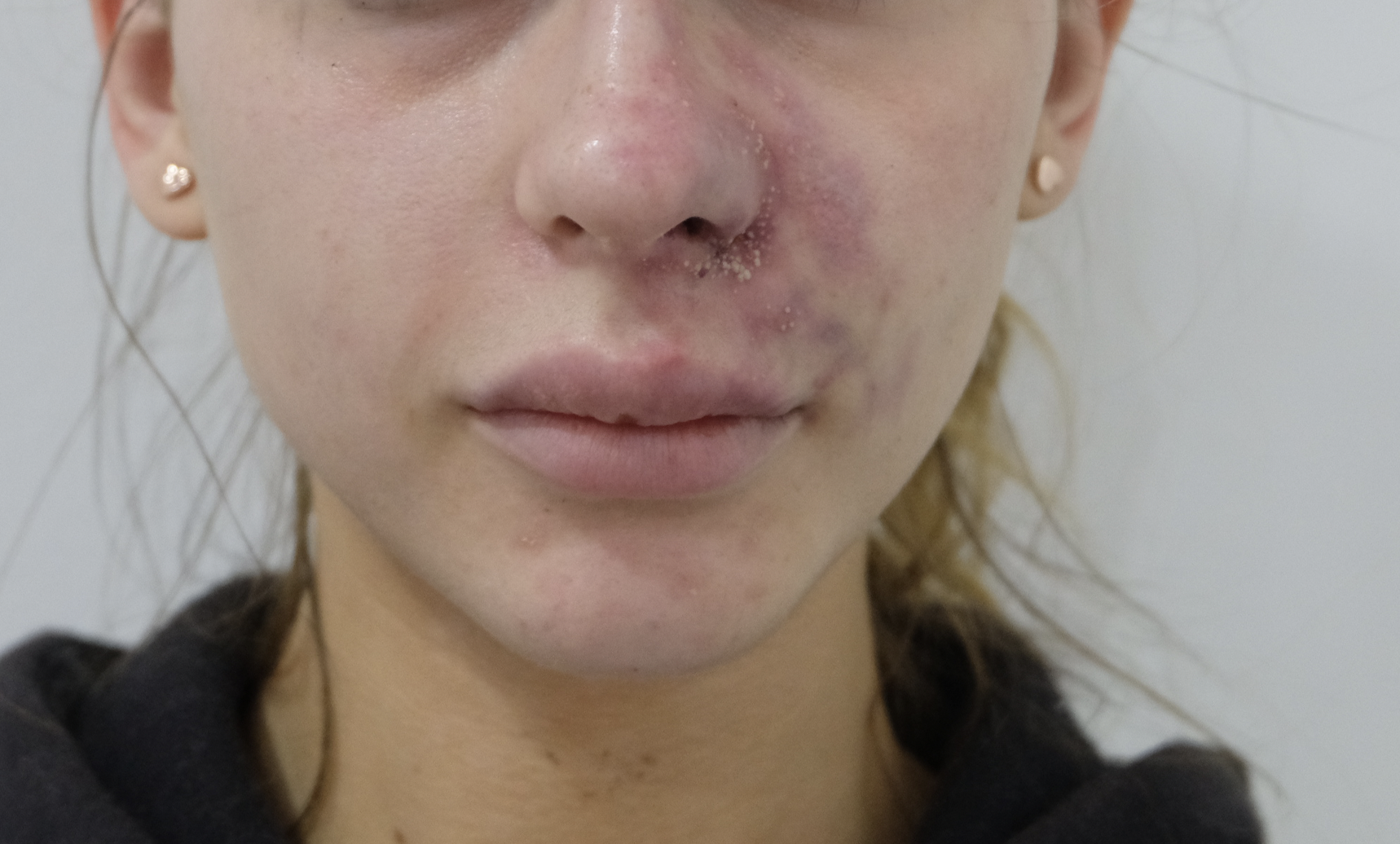
Image 1 · Presentation (24 h)
Reticular dusky discolouration of the left upper lip, philtrum and lateral nasal sidewall. Early pustulation at the alar base. Territory: superior labial artery with retrograde embolisation to the angular artery.
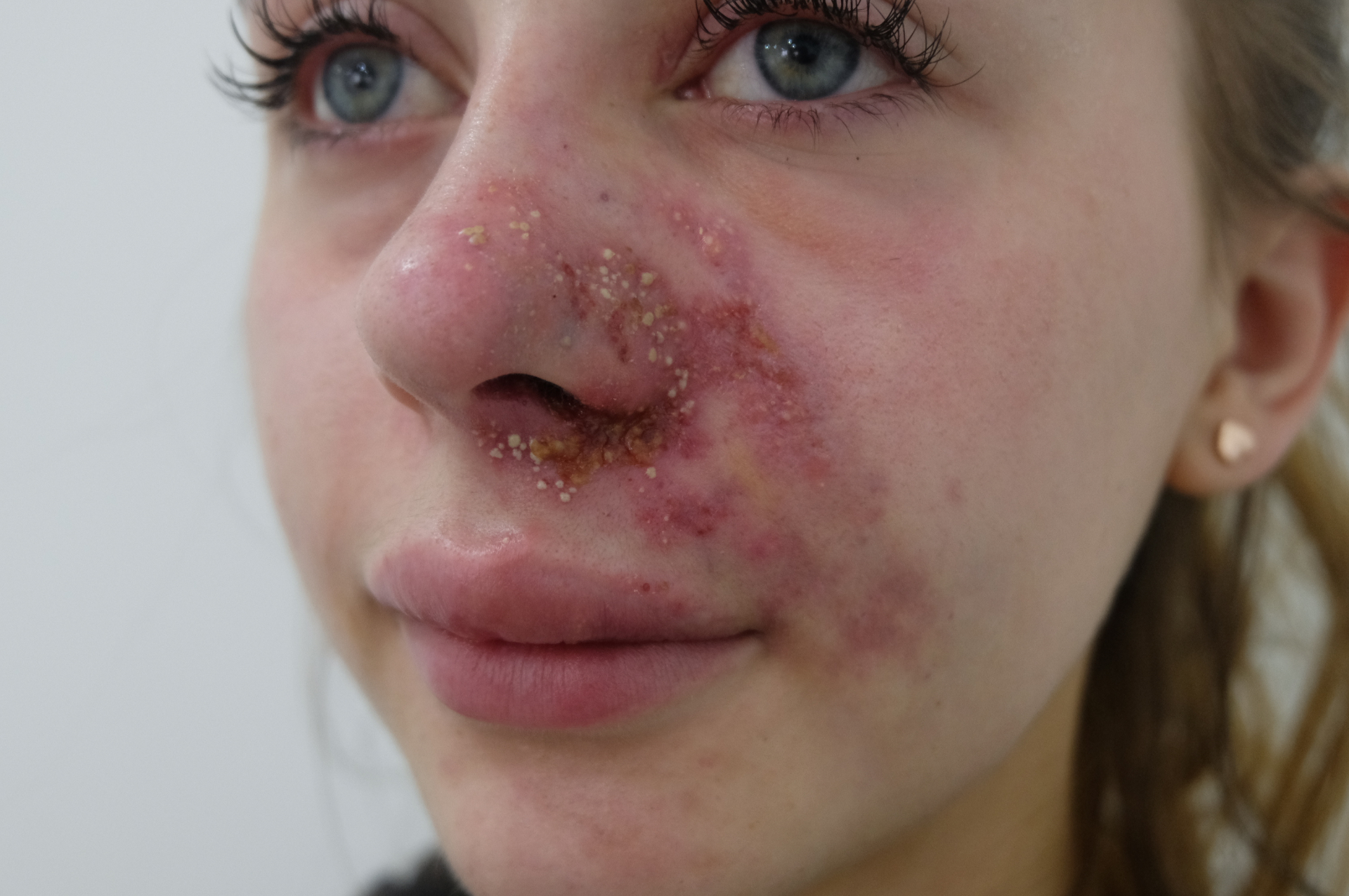
Image 2 · Day 1
Pustulation, crusting and superficial epidermal loss across the alar base, columella and lateral nasal sidewall — the expected evolution past the early hyaluronidase window. Underlying dermis remained viable.
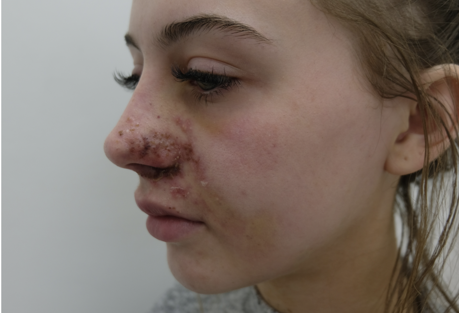
Image 3 · Day 3
Pustules consolidating into adherent crusts. Background erythema returning to previously dusky territory — clinical evidence of deep dermal reperfusion. Hyperbaric sessions ongoing.
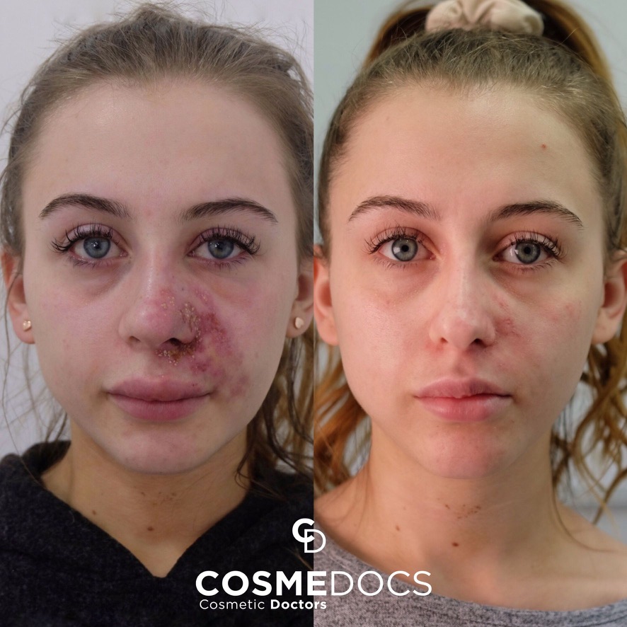
Image 4 · 3 months
Near-complete resolution. Residual fine post-inflammatory hyperpigmentation managed with tyrosinase inhibitors and photoprotection. No scarring requiring surgical revision.
7. Lessons for Clinic Design
The single largest determinant of vascular complication outcome is not the seniority of the injector. It is the written, rehearsed, in-date emergency protocol held by the clinic. Five operational requirements follow from the published evidence and from cases such as the one above.
(i) Stock. A minimum of 3000 units of hyaluronidase on-site at all times, with documented in-date status checked weekly. The DeLorenzi protocol can exhaust 1500 units in a single hour.
(ii) Pathways. A named local ophthalmology emergency contact, a named hyperbaric oxygen referral pathway, and a named acute hospital with a maxillofacial or plastics on-call rota, documented in the clinic emergency folder and known by every clinical staff member.
(iii) Drills. The vascular emergency protocol rehearsed in real time, with stopwatch, at least quarterly. Treat it as a fire drill: the first time it is performed should never be on a real patient.
(iv) Consent. Vascular occlusion, skin necrosis and the rare risk of visual loss explicitly named in the written consent for every filler treatment. The General Medical Council and the Joint Council for Cosmetic Practitioners both expect this as standard (JCCP, 2021).
(v) Audit. Every adverse event, including near-misses, documented and reviewed at clinic meetings. Pattern recognition across a clinic's case mix is the strongest internal mechanism for prevention.
8. Conclusion
Vascular complications in aesthetic practice are rare, recognisable and — with a complete, rehearsed protocol — survivable without permanent harm in the majority of cases. The published literature is unambiguous on the components of that protocol: high-dose pulsed hyaluronidase delivered early and locally, hyperbaric oxygen where indicated, anti-platelet and anti-inflammatory therapy, broad-spectrum antibiotic and antiviral cover, and immediate ophthalmology referral for any visual symptom. The variable that determines outcome is whether the clinic is ready to execute that protocol within the first hour. Readiness is a structural property of the clinic, not a personal property of the injector. Every aesthetic clinic operating in the United Kingdom should treat the construction, rehearsal and quarterly audit of a vascular emergency protocol as a non-negotiable condition of practice.
AI Disclosure
Large language model assistance was used during manuscript drafting and reference cross-checking. All clinical content, the case presentation and the final manuscript were reviewed and approved by named HSI Team clinical faculty, who retain full responsibility.
Competing Interests
The author(s) declare no competing financial or non-financial interests relevant to this work.
Funding
This work received no specific grant from any funding agency in the public, commercial, or not-for-profit sectors.
Ethics & Consent
Where applicable, ethical approval and informed patient consent were obtained in accordance with the Declaration of Helsinki. Reviews and commentaries did not require ethical approval.
HSI Editorial · Reflection & Forward Recommendations
Where we stand on this
Reflection
The single most important variable in vascular occlusion outcome is not the injector's skill at the moment the artery was struck — it is the clinic's readiness in the hour that followed. In the case shown, the patient presented at 24 hours, well past the textbook window, and still achieved full resolution. That outcome is not luck. It is the product of a written protocol, stocked hyaluronidase, a known route to hyperbaric oxygen, and a clinician willing to flood the territory rather than ration it.
Most aesthetic clinics in the UK are not set up for this. They carry one or two ampoules of hyaluronidase, no documented escalation pathway, and no rehearsed handover to a hyperbaric centre. The complication is therefore survivable in theory and disabling in practice.
Forward Recommendations
- Every injecting clinic should hold a minimum of 10,000 IU of hyaluronidase on site, written into the controlled-drug register, with expiry tracked monthly.
- Maintain a one-page vascular-occlusion protocol — recognition criteria, pulsed dosing schedule, named hyperbaric referral centre, on-call escalation number — displayed in every treatment room and rehearsed quarterly.
- Photograph and document every suspected vascular event from presentation through resolution. This is the dataset the field needs and currently lacks.
- Stop teaching 'do not use a needle in the lip'. Teach instead 'do not inject without a rehearsed first-hour protocol you can execute alone at 7pm on a Friday'. Readiness, not avoidance, is the safety strategy.
Editorial position of the Harley Street Institute. Authored by the HSI Clinical Review Board; not a substitute for the peer-reviewed evidence summarised above.
References
- Alam M, Kakar R, Dover JS, et al. Rates of vascular occlusion associated with using needles vs cannulas for filler injection: A systematic review and meta-analysis. JAMA Dermatology. 2021;157(2):174–180.
- Beleznay K, Carruthers JDA, Humphrey S, Jones D. Avoiding and treating blindness from fillers: a review of the world literature. Dermatologic Surgery. 2015;41(10):1097–1117.
- Beleznay K, Carruthers JDA, Humphrey S, Carruthers A, Jones D. Update on avoiding and treating blindness from fillers: a recent review of the world literature. Aesthetic Surgery Journal. 2019;39(6):662–674.
- Carey W, Weinkle S. Retraction of the plunger on a syringe of hyaluronic acid before injection: are we safe? Dermatologic Surgery. 2015;41 Suppl 1:S340–S346.
- Carruthers JDA, Fagien S, Rohrich RJ, Weinkle S, Carruthers A. Blindness caused by cosmetic filler injection: a review of cause and therapy. Plastic and Reconstructive Surgery. 2014;134(6):1197–1201.
- DeLorenzi C. New high dose pulsed hyaluronidase protocol for hyaluronic acid filler vascular adverse events. Aesthetic Surgery Journal. 2017;37(7):814–825.
- Goodman GJ, Magnusson MR, Callan P, et al. A consensus on minimizing the risk of hyaluronic acid embolic visual loss and suggestions for immediate bedside management. Aesthetic Surgery Journal. 2020;40(9):1009–1021.
- Hwang CJ, Mustak H, Gupta AA, Ramos RM, Goldberg RA, Duckwiler GR. Role of retrobulbar hyaluronidase in filler-associated blindness: evaluation of fundus perfusion and electroretinogram readings in an animal model. Ophthalmic Plastic and Reconstructive Surgery. 2018;35(1):33–37.
- Joint Council for Cosmetic Practitioners (JCCP). Clinical guidance for the prevention and management of complications associated with non-surgical cosmetic interventions. London: JCCP; 2021.
- Kapoor KM, Kapoor P, Heydenrych I, Bertossi D. Vision loss associated with hyaluronic acid fillers: a systematic review of literature. Aesthetic Plastic Surgery. 2020;44(3):929–944.
- Khan TT, Colon-Acevedo B, Mettu P, DeLorenzi C, Woodward JA. An anatomical analysis of the supratrochlear artery: considerations in facial filler injections and preventing vision loss. Aesthetic Surgery Journal. 2018;37(2):203–208.
- Sotorra-Figuerola D, Sanchez-Torres A, Valmaseda-Castellón E, Gay-Escoda C. Hyperbaric oxygen therapy for the management of dermal filler-induced vascular compromise: a systematic review. Journal of Cosmetic Dermatology. 2020;19(11):2814–2820.
- Urdiales-Gálvez F, Delgado NE, Figueiredo V, et al. Treatment of soft tissue filler complications: expert consensus recommendations. Aesthetic Plastic Surgery. 2018;42(2):498–510.
- van Loghem JAJ, Humzah D, Kerscher M. Cannula versus sharp needle for placement of soft tissue fillers: an observational cadaver study. Aesthetic Surgery Journal. 2018;38(1):73–88.
- van Loghem J, Yutskovskaya YA, Philip Werschler W. Calcium hydroxylapatite: over a decade of clinical experience. Journal of Clinical and Aesthetic Dermatology. 2015;8(1):38–49.
Declarations
- Peer review:
- This article underwent single-blind external peer review by at least two independent reviewers, followed by editorial acceptance.
- Conflicts of interest:
- The author(s) declare no competing financial or commercial interests relating to the content of this article. Editorial decisions are made independently of the Harley Street Institute's commercial training activities.
- Funding:
- No external funding was received for the preparation of this article.
- Licence:
- © 2026 Harley Street Institute. Open access article distributed under the Creative Commons Attribution 4.0 International Licence (CC BY 4.0), permitting unrestricted use with appropriate citation.
Post-publication review
Discuss this article with the journal AI
Ask a clinical question about this article, or flag a possible error. Our AI agent will reply in real time, log your input, and — if you have identified a credible mistake — escalate it to the HSI editorial team for review and a published correction notice.
AI responses are generated by an assistant model. They do not constitute medical advice. Editorial corrections are only applied after a named HSI editor reviews and signs off.
Hi. I'm the Aesthetic Intelligence reader AI for this article.
Try: "Explain section 3 in plain English." · "What's the evidence for the 1-in-6,410 figure?" · "How does this compare to UK NICE guidance?"