
The Masseter Reconsidered
Functional anatomy, architecture and bone interaction for the aesthetic practitioner.
Abstract
Background. The masseter is routinely taught to aesthetic practitioners as a two-part elevator of the mandible. That simplification does not account for paradoxical bulging after botulinum toxin, compensatory hypertrophy of untreated regions, smile asymmetry from spread into the risorius, or patients whose lower-face width is skeletal rather than muscular.
Methods. Narrative review and educational synthesis of cadaveric dissection studies, muscle architecture and physiological cross-sectional area measurements, myosin heavy-chain fibre-typing work, biomechanical and electromyographic studies of jaw closing, fascial and risorius anatomy, and human computed tomography and cone-beam data on mandibular bone following masseter neuromodulation.
Results. The masseter is multipennate and multilaminar, comprising superficial, intermediate and deep parts separated by aponeurotic laminae, with a separately described coronoid part and a deep inferior tendon partitioning superficial from sub-tendinous contractile tissue. Its fibre population is dominated by large, slow, oxidative and hybrid fibres retaining fetal and alpha-cardiac myosin isoforms. Leverage and cross-sectional size make it the dominant jaw elevator, although the share attributed to each elevator varies with model and task. The parotideomasseteric fascia is a single sheet continuous with the parotid capsule, and the risorius arises from fascia in its plane in variable patterns. Twelve-month placebo-controlled computed tomography found no difference in bigonial width, cortical thickness, flare angle, gonial angle or bone density at cosmetic doses, while smaller studies report modest trabecular change.
Conclusion. Layer-aware assessment, intramuscular placement away from the anterior border, individualised dosing and explicit separation of muscular from skeletal width improve safety and expectation setting in masseter treatment. Neuromodulation reduces muscular prominence but does not remodel a flared mandibular angle.
Postgraduate Level
Complete this article to earn your certificate
Read the article, complete a short assessment, and submit your reflection to receive your AiCE Points certificate. Equivalence to 1 CPD/CME point — we do not award CPD/CME directly.
Learning Objectives
- 1Describing the multipennate, multilaminar architecture of the masseter, including the deep inferior tendon and coronoid part
- 2Explaining how physiological cross-sectional area and pennation angle determine force rather than visible bulk
- 3Relating the slow, hybrid fibre-type profile to endurance, parafunction and response to neuromodulation
- 4Using the parotideomasseteric fascia and variable risorius origin to reduce smile-related complications
- 5Distinguishing muscular from skeletal lower-face width and interpreting the mandibular bone evidence after masseter toxin
The masseter is not a two-layer block. It is a multipennate, multilaminar force system whose internal tendons, variable fibre orientations and close fascial relationship to the risorius directly influence assessment, treatment planning and complications. The key clinical distinction is equally simple: define how much lower-face width is muscle, bone and soft tissue before promising a change.
1. Introduction
Every aesthetic practitioner who treats “square jaw”, bruxism-related pain or lower-face width injects the masseter. Few have been taught it as anything more than “superficial part, deep part, elevates the mandible”. That simplification has consequences: paradoxical bulging after treatment, compensatory hypertrophy of untreated regions, smile asymmetry from toxin spread into the risorius, and unrealistic patient expectations where the width is skeletal rather than muscular.
Understanding the masseter properly means understanding four things: how it is built (layers and pennation), what it is made of (fibre types and metabolism), how it acts (torque, leverage and its share of jaw-closing force), and what it does to the bone beneath it over years of loading. This article addresses each in turn.
2. Gross Anatomy: A Working Summary
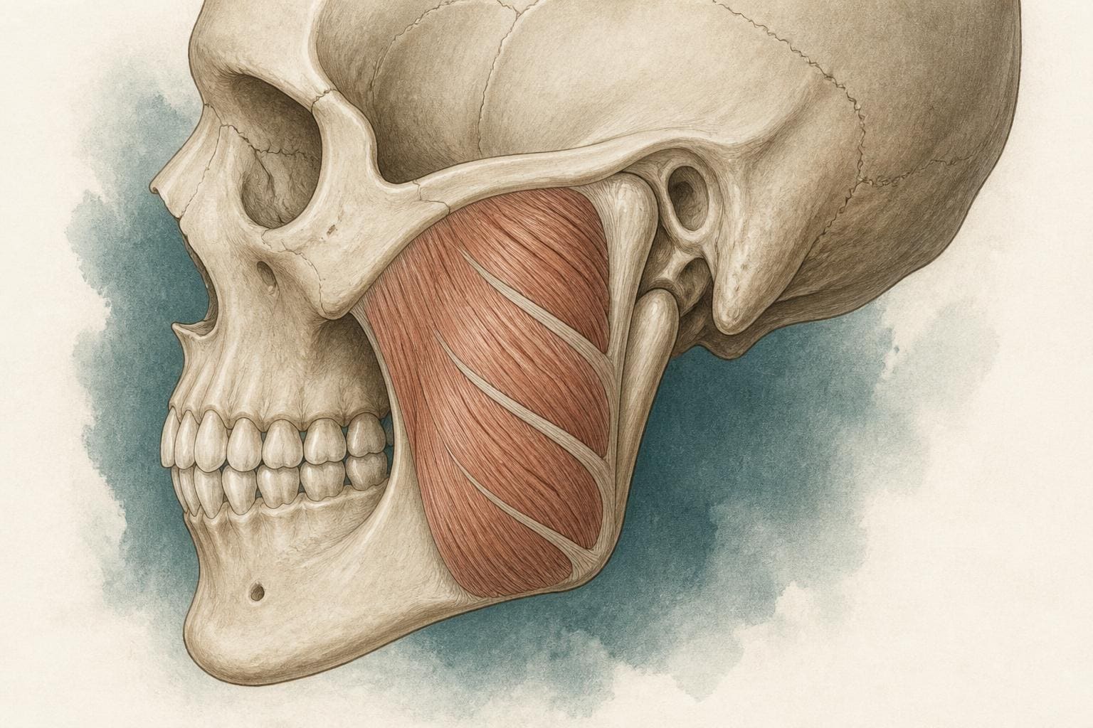
The masseter is a thick, quadrilateral muscle spanning from the zygomatic arch to the lateral surface of the mandibular ramus and angle. Its superficial part arises by a strong aponeurosis from the maxillary process of the zygomatic bone and the anterior two-thirds of the inferior border of the zygomatic arch, running inferiorly and posteriorly at roughly 60 degrees to the occlusal plane to insert on the angle and lower half of the ramus (the masseteric tuberosity). The deep part arises from the deep surface and posterior third of the arch, runs more vertically, and inserts on the upper ramus and toward the coronoid process. The two parts are fused anteriorly and separated posteriorly, where the deep part is partly visible above the superficial part beneath the parotid.
Innervation is from the masseteric nerve, a branch of the anterior division of the mandibular nerve (V3), which passes laterally through the mandibular notch with the masseteric artery (from the maxillary artery) to enter the deep surface of the muscle. This is worth stating plainly: the masseter is a trigeminal muscle, embryologically a first-pharyngeal-arch structure, and therefore developmentally and neurologically unrelated to the facial-nerve muscles of expression that lie on top of it.
Superficial relations are the ones that matter to the injector: the parotid gland (overlapping the posterior third), the parotid duct crossing the muscle roughly a finger’s breadth below the arch, the transverse facial artery, the buccal and marginal mandibular branches of the facial nerve running on the muscle’s fascia deep to the SMAS, the risorius and zygomaticus muscles anteriorly, and the buccal fat pad at the anterior border. Deep to the muscle lie the ramus, the mandibular notch and, medially through the notch, the temporalis tendon and the lateral pterygoid.
3. Layered Architecture: The Feather Within the Block
Where this is on the face
The schematic beside this is a slice through the shaded area: the muscle running from the cheekbone down to the angle of the jaw, seen from the side with the skin removed.
3.1 From two layers to three (and a coronoid part)
The two-layer textbook model was overturned in 2000 by Gaudy and colleagues, who dissected and imaged the jaw elevators in 169 cadavers. They described the masseter as a multipennate muscle built from a succession of alternating muscle and aponeurotic sheets, organised into three well-defined parts. The superficial masseter comprised two alternating musculo-aponeurotic layers oriented at about 60 degrees to the occlusal plane; the intermediate masseter a single layer at about 90 degrees; and the deep masseter three layers, with the outer laminae at roughly 90 degrees and a middle lamina at about 110 degrees.
The picture has been refined further. In 2021 Mezey and co-workers reported a consistently identifiable third, deep layer running from the medial surface of the zygomatic process of the temporal bone to the root and posterior margin of the coronoid process — a “coronoid part” positioned to stabilise and retract the anterosuperior mandible rather than to elevate it. And in 2017 Lee and colleagues, investigating paradoxical bulging after botulinum toxin, showed that the superficial part is itself lamellar: a superficial muscle belly, then a broad, deep inferior tendon (DIT) attached to the lower third of the ramus and the inferior mandibular border, then a further thin muscle belly beneath the tendon, and finally the insertions of the middle and deep parts on periosteum.
The practical result is a muscle that, in cross-section, resembles a stack of feathers pressed together: muscle fibres run obliquely between tendinous laminae that themselves run from bone to bone. This is the “feather-shaped” (pennate) architecture the article title alludes to, and it is the single most important concept for understanding masseteric force.
| Part | Typical fibre direction | Principal mechanical role | Clinical relevance |
|---|---|---|---|
| Superficial | Upward and forward; oblique | Elevation and protrusion | Dominant visible lower-masseter bulk; deep inferior tendon may partition the belly. |
| Intermediate | More vertical | Elevation | May remain active when treatment is concentrated superficially. |
| Deep | Near vertical to slightly posterior | Elevation, retrusion and stabilisation | Posterior and deeper position changes both vector and accessibility. |
| Coronoid part | Toward coronoid root and posterior margin | Probable stabilisation and retrusive contribution | Anatomically consistent but not a routine surface landmark. |
3.2 Why pennation matters
A parallel-fibred muscle (think sartorius) has long fibres running the muscle’s whole length. Its force is proportional to its anatomical cross-section, and it can shorten over a large range. A pennate muscle sacrifices excursion for force: by angling short fibres onto a central tendon, it packs many more fibres into the same volume. The relevant measure is physiological cross-sectional area (PCSA): muscle volume divided by fibre length, corrected by the cosine of the pennation angle. PCSA, not visible bulk, predicts maximal isometric force.
Van Eijden, Korfage and Brugman (1997) measured this directly in eight cadavers. Relative to the jaw openers, the jaw closers — superficial and deep masseter, anterior and posterior temporalis, medial pterygoid — had shorter sarcomeres at the closed-jaw position, larger masses of contractile and tendinous tissue, larger PCSAs, larger pennation angles, shorter fibre lengths, shorter moment arms and lower fibre-length-to-muscle-length ratios. The jaw closers are, in other words, force-optimised; the openers are velocity- and displacement-optimised.
Fibre lengths in the masseter are in the order of 2–3 cm in a muscle 6–7 cm long; pennation angles reach 15–25 degrees in the superficial part. The consequence: a muscle roughly the size of a palm can generate several hundred newtons per side, but only over a shortening range of a centimetre or so — exactly what the mandible needs at the end of the closing stroke, where the teeth meet the bolus.
Because force is distributed across multiple laminae with different fibre orientations, weakening one lamina — the superficial belly, where most injectors deliver toxin — leaves the others free to contract and, over repeated cycles, to hypertrophy. This architecture provides a plausible anatomical basis for paradoxical masseteric bulging after toxin treatment and for compensatory prominence reported in untreated regions. The practical response is layer-aware assessment, conservative placement and follow-up; ultrasound may help identify the deep inferior tendon, muscle thickness and the location of a focal residual bulge.
4. Fibre Types and Energetics: A Muscle That Reads Like Heart and Fetus
4.1 An atypical myosin profile
Limb muscle contains pure type I (slow oxidative), IIA (fast oxidative-glycolytic) and IIX (fast glycolytic) fibres in a mosaic, with fast fibres typically larger than slow. The masseter breaks almost every one of these rules.
Immunohistochemical and biochemical work since the 1980s (Ringqvist; Butler-Browne; Korfage and van Eijden; Österlund and colleagues) has shown that the masseter, temporalis and pterygoids contain an unusually large proportion of hybrid fibres expressing more than one myosin heavy-chain (MyHC) isoform; that many fibres express not only MyHC-I but also fetal (neonatal) MyHC and alpha-cardiac MyHC — isoforms essentially absent from adult limb muscle; and that, uniquely, type I fibres are larger in cross-section than type II fibres. Type I fibres are generally the most abundant class, with proportions varying markedly between individuals and between regions of the muscle. Type II subclasses in the masseter are small, and IIB-type MyHC transcripts, which are not expressed in adult human limb muscle, have been found abundantly in a subset of masseter fibres.
Fibre-type composition also changes through development in a pattern distinct from limb muscle: the young masseter contains type I, IM, IIC, IIAB and IIB fibres with only scarce IIA and with fetal MyHC persisting, and the growth pattern of fibre diameters between childhood and adulthood follows a course unique to the jaw muscles, presumably tracking eruption and the maturation of mastication and speech.
4.2 What this means functionally
Hybrid fibres have contractile properties intermediate between their constituent isoforms. A muscle rich in I/IIA hybrids has a smooth, finely graded velocity-force spectrum rather than a two-speed gearbox. Combined with the predominance of large, slow, oxidative type I fibres and a rich capillary bed, this makes the masseter a fatigue-resistant, endurance muscle that can hold tonic postural jaw position all day, sustain long chewing sequences and — critically for the aesthetic clinic — sustain hours of nocturnal clenching without failing. The metabolic profile is predominantly oxidative: high mitochondrial density, reliance on aerobic phosphorylation, with the small fast fibres providing the brief anaerobic bursts needed for a hard bite.
The retention of developmental and cardiac isoforms is thought to reflect the masseter’s origin from first-arch (branchiomeric) mesoderm rather than somitic mesoderm, and to underpin its distinct response to load, denervation and ageing.
A slow, oxidative, hybrid-rich muscle is precisely the phenotype that hypertrophies under chronic low-frequency, high-duration load — the bruxism pattern — and that recovers function after neuromodulation in a graded way. It may also contribute to inter-individual variability in functional recovery after botulinum toxin. Dose and retreatment interval, however, should be individualised by product labelling, muscle size, indication, response and adverse effects rather than inferred from fibre type alone.
5. Torque, Leverage and the Masseter’s Share of Bite Force
5.1 The mandible as a class III lever
For most bite points the mandible functions as a class III lever: the fulcrum is the temporomandibular joint, the effort (muscle) is applied between the fulcrum and the load (tooth). The mechanical advantage of each elevator is the ratio of its moment arm about the condyle to the bite-point moment arm. Two consequences follow. First, force at the molars is always greater than at the incisors, because the load arm is shorter. Second, a muscle inserting further from the condyle and with a more vertical line of action has better leverage.
The masseter scores well on both. Skeletal studies measuring adductor leverage across the tooth row find leverage greatest for the masseter, then the anterior temporalis, then the medial pterygoid, with the advantage increasing at posterior bite points. Cephalometric and sectional work by Hannam and Wood showed the masseter was always a more efficient producer of vertically oriented bite force than the medial pterygoid, being roughly two-thirds larger in cross-section and inclined more anteriorly relative to the occlusal plane. The same authors, measuring unilateral first-molar bite force directly (mean 189 ± 78 N in their sample), found that masseter and medial pterygoid cross-sectional size correlated significantly with bite force, whereas lever-arm length did not.
5.2 The percentage question
Biomechanical models consistently identify the masseter, temporalis and medial pterygoid as the principal contributors to jaw closing, but the percentage assigned to each muscle varies with model assumptions, bite point, mandibular position, PCSA estimates and activation strategy. A commonly repeated 43/36/21 split should therefore be treated as an illustrative model estimate, not a universal physiological constant. The lateral pterygoid’s superior head contributes to the closing stroke by seating the condyle-disc complex rather than by elevation, and is conventionally omitted from these figures. Musculoskeletal simulations of maximal biting likewise identify the superficial masseter, anterior temporalis and medial pterygoid as the dominant contributors, with working-side muscles producing more force than non-working-side muscles in unilateral biting.
These percentages describe force capacity, not activation. Electromyography during chewing shows the masseter and temporalis firing in a task-dependent ratio: the temporalis dominates in positioning and in the early closing phase, the masseter in the power stroke as the teeth approach and pass through occlusion. Superficial and deep masseter are also not synchronous — the deep part activates earlier and contributes a retrusive, condyle-stabilising vector; the superficial part delivers the protrusive-elevating power stroke.
5.3 Direction, not just magnitude
The superficial masseter’s line of action runs upward and forward (roughly 60 degrees to the occlusal plane), so it elevates and protrudes. The deep part runs almost vertically with a slight backward inclination, so it elevates and retrudes, and in unilateral chewing helps swing the working-side condyle. The coronoid part described by Mezey acts on the anterosuperior mandible, plausibly stabilising the coronoid during the temporalis-driven closing stroke. The masseter is therefore not one torque but a set of partially independent torques applied around the condylar axis — and a hypertrophied superficial part will change the shape of the angle differently from a hypertrophied deep part.
Weakening the masseter can reduce bite-force capacity and may lessen symptoms in selected patients, although the effect varies and treatment of bruxism or myofascial pain should sit within a broader diagnostic and dental plan. It is also the reason patients may report chewing fatigue with tough foods for some weeks: the remaining muscles have to compensate, and the temporalis — the next largest contributor — may hypertrophy visibly at the temporal fossa in long-term, high-dose patients.
6. The Parotideomasseteric Fascia: Why One Name for Two Structures
6.1 A single sheet, not a fusion
Students often ask why the fascia over the masseter is called the “parotideomasseteric” (or parotid-masseteric) fascia, as if two fasciae had combined. The answer is that it is one continuous sheet that happens to cover two organs. The investing layer of deep cervical fascia, ascending from the neck, splits to enclose the parotid gland in a dense capsule; its superficial lamina then continues upward and forward over the lateral surface of the masseter to attach to the zygomatic arch. Over the gland it is the parotid capsule; over the muscle it is the masseteric fascia; the composite name simply acknowledges that no anatomical boundary separates them. The gland is moulded onto the posterior part of the muscle inside this shared compartment, and the parotid duct exits the gland and crosses the muscle beneath the same sheet before piercing the buccinator.
Over the masseter this fascia is thin but very strong, firmly attached to the arch above and the inferior mandibular border below, and continuous posteriorly with the fascia of the sternocleidomastoid. Its deep surface adheres to the epimysium of the superficial masseter; its superficial surface is separated from the SMAS by a loose areolar plane in which the facial nerve branches run.
6.2 Relationship to the SMAS and the retaining ligaments
The SMAS is a separate, more superficial layer — the facial continuation of the platysma sheet — and the parotideomasseteric fascia is the deep fascia beneath it. The two are tethered by the masseteric cutaneous ligaments (Furnas), a series of fibrous septa passing from the anterior border of the masseter through the SMAS to dermis. These ligaments are why the anterior masseteric border is a landmark for jowl formation and why sub-SMAS dissection over the masseter requires deliberate release. For the injector, the significance is different: toxin injected superficially can pool in the areolar sub-SMAS plane above the fascia and reach the mimetic muscles, whereas placement within the muscle reduces exposure of superficial mimetic muscles but does not eliminate diffusion, which remains influenced by dose, concentration, volume and local anatomy.
| Structure / plane | Why it matters | Risk-control principle |
|---|---|---|
| Risorius and zygomatic muscles | Spread can weaken commissure movement and produce smile asymmetry. | Keep away from the anterior border; reassess individual anatomy and depth. |
| Parotid gland and duct | Posterior overlap and duct course vary between patients. | Do not rely on one surface line; use ultrasound when boundaries are uncertain. |
| Facial nerve branches | Branches traverse the superficial fascial environment. | Intramuscular placement and conservative volume reduce unintended exposure. |
| Deep inferior tendon | Can separate superficial and sub-tendinous contractile tissue. | Consider when focal paradoxical bulging persists after an apparently adequate treatment. |
6.3 How the fascia and the risorius develop together
The muscles of facial expression derive from second-arch mesoderm that migrates superficially across the face as a continuous sheet — the primitive platysma or panniculus carnosus — and subsequently differentiates into individual muscles. The SMAS is the fibrous remnant of that sheet where muscle fibres did not persist. The risorius is best understood as a detached lateral bundle of this platysma sheet that has kept its muscle fibres: a phylogenetically recent, inconstant muscle that has no bony origin at all. It therefore takes its origin from whatever fibrous tissue lies in its plane — the SMAS, the parotideomasseteric fascia, or the masseter’s own tendon. This developmental origin is the answer to “why is the risorius combined with the fascia”: the fascia is the muscle’s origin because there is nothing else in that plane for it to arise from.
7. The Risorius: Variable, Inconstant and Clinically Decisive
7.1 Origin patterns
Bae and colleagues (2014), dissecting 46 hemifaces to resolve the confusion about the risorius’s plane, found three patterns. In 58.7% the muscle arose solely from the fascial layer superficial to the SMAS, in the same plane as the platysma. In 6.5% it arose solely from the masseter tendon. In 34.8% it arose from fascial layers both superficial and deep to the SMAS — that is, from both the SMAS and the parotideomasseteric fascia. The muscle is absent altogether in a meaningful minority of individuals and highly variable in width and thickness where present. It passes horizontally forward, superficial to the platysma and buccinator, to insert into the modiolus at the angle of the mouth, and is supplied by buccal branches of the facial nerve.
7.2 Function
The risorius retracts the oral commissure laterally without elevating it — the “horizontal” or closed-mouth smile, and a component of the grimace. Its contribution to smile aesthetics is modest compared with zygomaticus major, but its loss on one side is conspicuous because the commissure then drifts toward the unaffected side on smiling.
7.3 Why it matters for masseter injection
Because the risorius arises from the fascia overlying the anterior-superior masseter, and because zygomaticus major and minor course across the muscle’s anterior border, toxin diffusing forward or superficially from the injection point reaches facial-nerve muscles that were never the target. The published pattern of masseter-related complications — asymmetric smile, commissure retraction weakness, “sunken” cheek from combined risorius and buccinator effect — maps directly onto this anatomy. Common techniques favour conservative intramuscular placement within the palpated lower masseter and away from the anterior border, but no drawn surface box is universally safe. The parotid duct, gland overlap, facial vessels, risorius and individual muscle borders vary; ultrasound is especially valuable where anatomy is uncertain, muscle is thin or prior treatment has altered the contour.
8. Hypertrophy and the Mandible: What the Bone Actually Does
8.1 Bone follows load
Wolff’s law predicts that bone adapts to chronic loading, and the masseteric attachment is a textbook example. Chronic clenching and grinding increase bone density at muscle attachment sites and remodel trabecular architecture in the angle and ramus. Over years, the masseteric tuberosity becomes more rugose, the gonial angle more acute, and — most importantly for the aesthetic assessment — the angle itself may flare laterally and evert, producing a bony contribution to the square lower face that no muscle treatment will address. The Korean and Japanese contouring literature has long distinguished “muscular” from “bony” square jaw on this basis, and a substantial proportion of patients presenting with masseteric prominence have both.
Developmentally, the direction of causation runs both ways: masseter size correlates with bigonial width, intergonial flare and a shorter, more horizontal ramus, and morphometric studies associate high putative molar bite force with a face having large intergonial width, small intercondylar width, a narrow dental arch and a forward maxilla and mandible. Muscle and bone co-develop as a single loading system.
8.2 Does reducing the muscle reverse the bone?
This is where the evidence is more nuanced than either enthusiasts or sceptics suggest.
Short-term human CT. In a retrospective series of ten women scanned before and three months after standardised bilateral masseter toxin injection, soft-tissue volume fell but mandibular cortical thickness, bone thickness and volume did not change. The authors explicitly concluded that the popular idea of mandibular flaring “resolving” once the muscle is partially paralysed was not supported, and that patients with both hypertrophy and bony flaring will usually need a combined muscular and skeletal approach.
Twelve-month randomised data. A large placebo-controlled onabotulinumtoxinA programme for masseter muscle prominence used serial multidetector CT read by dental radiologists. Quantitative assessment of bigonial width, cortical thickness under the masseteric insertion, mandibular flare angle and gonial angle found no difference between treated (n = 150) and placebo (n = 37) after one or two treatment cycles at 48–96 units, and a companion analysis found no negative effect on mandibular bone density over the year. Isolated subclinical radiographic changes occurred in all groups including placebo and were adjudicated as artefact. The authors appropriately note that effects beyond twelve months cannot be excluded.
Signals of change. Against this, a prospective CBCT trial reported reduced bone volume at the gonial angle at six months in participants treated twice within four months but not in those treated once; a small series reported reduced trabecular density six to ten weeks post-treatment; and a 2025 matched-pair panoramic-radiograph study in 42 bruxism patients found statistically significant reductions in trabecular fractal dimension specifically in the angulus region six months after 50 units per masseter, although post-treatment values did not differ from untreated controls — implying regression toward normal rather than pathological loss. Rodent studies consistently show condylar and alveolar bone loss after masseter toxin, but rodent jaw bone turnover is far faster and the doses are not comparable.
Synthesis. The best current reading is that (i) chronic hypertrophic loading densifies and remodels the angle; (ii) unloading the muscle with neuromodulator produces measurable but modest trabecular normalisation at the angulus, most detectable in bruxers with elevated baseline density; (iii) macroscopic mandibular morphology — width, flare, gonial angle, cortical thickness — does not change over twelve months at cosmetic doses; and (iv) there is no evidence of clinically harmful bone loss in adults at standard dosing, though long-term, high-frequency data are lacking and caution is warranted in adolescents and in patients with compromised bone health.
| Evidence window | Signal reported | Interpretation |
|---|---|---|
| 3-month human CT | Soft-tissue reduction without clear cortical thickness, bone thickness or volume change. | Muscle reduction should not be presented as reversal of skeletal flare. |
| 12-month placebo-controlled CT | No treatment-placebo difference in bigonial width, cortical thickness, flare angle, gonial angle or bone density. | Reassuring at studied adult doses and intervals; longer-term exposure remains less certain. |
| Smaller CBCT / radiographic studies | Some trabecular or regional changes, particularly after repeated treatment or in bruxism cohorts. | Potential remodelling signal; methods and clinical significance are heterogeneous. |
| Animal studies | Consistent unloading-related bone effects. | Biological plausibility, but dose scaling and turnover limit direct translation to adults. |
Assess before you treat. Palpate the angle at rest and during clench; a prominence that persists at rest, feels hard, and is confirmed on panoramic radiograph or CBCT is skeletal. Counsel accordingly: neuromodulator will narrow the muscular component within three months but will not slim a flared angle. Where a flared mandibular angle is the dominant cause of width, set that expectation before treatment and consider maxillofacial assessment if the patient seeks skeletal change. Treat the overlying soft-tissue envelope only when a separate indication is present.
9. Translating the Anatomy into Technique
Bringing the sections together, the anatomically literate approach to the masseter looks like this:
- Map the muscle, not the cheek. Identify the anterior and posterior borders on clench, the inferior mandibular border and the likely parotid gland and duct territory. Marking defines the intended muscle field; it does not create an anatomically guaranteed safe quadrilateral.
- Think in layers. The superficial belly, the deep inferior tendon and the sub-tendinous belly all lie within the lower third. Use the lowest-complexity pattern that addresses the palpable or sonographic target. Focal residual bulging after treatment should prompt reassessment of depth and tendon anatomy before additional placement.
- Respect the fascia. Confirm intramuscular placement. Superficial deposition increases exposure of the sub-SMAS plane and adjacent mimetic muscles; depth reduces but does not abolish diffusion risk.
- Dose to the muscle you have. Thickness on ultrasound, degree of parafunction and treatment history all change the requirement. Individualise treatment to muscle thickness, indication, product, prior response and functional risk; fibre-type theory does not determine a clinical dose.
- Anticipate compensation. Warn patients about transient chewing fatigue and about the temporalis. Examine the temporal fossa at every review.
- Separate muscle from bone. Where the width is skeletal, say so, and offer a combined plan.
- Use ultrasound. Layer identification, thickness measurement and real-time needle placement convert the anatomy above from theory into reproducible practice.
10. Conclusion
The masseter is not a block; it is a stack of feathers. Its multipennate, multilaminar architecture explains both its extraordinary force-to-size ratio and its capacity to bulge paradoxically when only one lamina is treated. Its slow, hybrid, developmentally unusual fibre profile explains its endurance, its response to chronic parafunction and its variable response to neuromodulation. Its leverage and line of action make it the dominant jaw elevator, a major contributor to closing capacity, with its exact share dependent on the model and task. The fascia that covers it is a single sheet shared with the parotid, from which the risorius — a platysmal remnant with no bony origin — takes its variable origin, placing smile function within millimetres of the injection field. And the bone beneath it remodels under decades of load in ways that neuromodulator normalises at the trabecular level but does not reverse at the level of shape.
Practitioners who internalise these five ideas will treat the masseter more safely, more predictably and with better-counselled patients. The anatomy is not difficult. It is simply more detailed than it is usually taught.
Editorial and clinical notice
This narrative review is educational and is intended for appropriately qualified healthcare professionals. It does not replace hands-on training, product-specific prescribing information, local regulation, individual anatomical assessment, consent or clinical judgement. The article was edited with AI assistance and reviewed for publication by the author.
Copyright 2026 Dr Ahmed Haq and Harley Street Institute. Published in Aesthetic Intelligence under CC BY 4.0. ISSN 2979-8116 (Online).
References
Reference details were standardised from the supplied manuscript and checked against key indexed records. DOI and pagination should receive a final production check before external journal submission.
- Gaudy JF, Zouaoui A, Bravetti P, Charrier JL, Guettaf A. Functional organization of the human masseter muscle. Surg Radiol Anat. 2000;22(4):181–190. View source
- Mezey SE, Müller-Gerbl M, Toranelli M, Türp JC. The human masseter muscle revisited: first description of its coronoid part. Ann Anat. 2022;240:151879. View source
- Lee HJ, Kang IW, Won SY, et al. The anatomical basis of paradoxical masseteric bulging after botulinum neurotoxin type A injection. Toxins. 2017;9(1):14. View source
- Compensatory masseteric bulging: a novel observation and its implications for botulinum neurotoxin injection techniques. J Cosmet Dermatol. 2025. View source
- Van Eijden TMGJ, Korfage JAM, Brugman P. Architecture of the human jaw-closing and jaw-opening muscles. Anat Rec. 1997;248(3):464–474. View source
- Korfage JAM, van Eijden TMGJ. Differences in myosin heavy-chain composition between human jaw-closing muscles and supra- and infrahyoid muscles. Arch Oral Biol. 2001. View source
- Ringqvist M. Histochemical enzyme profiles of fibres in human masseter muscles with special regard to fibres with intermediate myofibrillar ATPase reaction. J Neurol Sci. 1973;18(2):133–141.
- Österlund C, et al. Differences in fibre type composition between human masseter and biceps muscles in young and adults reveal unique masseter fibre type growth pattern. Anat Rec. 2011;294(7):1158–1169. View source
- Horton MJ, et al. Abundant expression of myosin heavy-chain IIB RNA in a subset of human masseter muscle fibres. Arch Oral Biol. 2001. View source
- Korfage JAM, van Eijden TMGJ. Regional differences in fibre type composition in the human temporalis muscle. J Anat. 1999;194:355–362. View source
- Hannam AG, Wood WW. Relationships between the size and spatial morphology of human masseter and medial pterygoid muscles, the craniofacial skeleton, and jaw biomechanics. Am J Phys Anthropol. 1989;80(4):429–445. View source
- Relationships between the size, position, and angulation of human jaw muscles and unilateral first molar bite force. J Dent Res. 1989. View source
- The ontogeny of maximum bite force in humans. J Anat. 2020. View source
- The effective way of botulinum toxin injection to reduce bite force: preliminary study. 2025. View source
- Muscle and joint mechanics during maximum force biting following total temporomandibular joint replacement surgery. Biomech Model Mechanobiol. 2024. View source
- Bae JH, Lee JG, Youn KH, et al. Surgical consideration of the anatomic origin of the risorius in relation to facial planes. Aesthet Surg J. 2014;34(7):NP43–NP49. View source
- Chang CS, Bergeron L, Yu CC, Chen PKT, Chen YR. Mandible changes evaluated by computed tomography following botulinum toxin A injections in square-faced patients. Aesthetic Plast Surg. 2011;35:452–455. View source
- Longitudinal computed tomographic evaluation of mandibular bone morphology following onabotulinumtoxinA treatment of masseter muscle prominence: results of a 12-month, repeat-treatment, placebo-controlled study. Aesthet Surg J. 2026. View source
- Longitudinal computed tomography indicates no negative impact of onabotulinumtoxinA on mandibular bone density in a 12-month, double-blind, randomized, placebo-controlled study. Aesthet Surg J. 2025. View source
- Reduction of masseter muscle prominence after treatment with onabotulinumtoxinA: primary results from a randomized phase 2 study. J Am Acad Dermatol. 2024. View source
- Is mandibular cortical bone and trabecular microarchitecture altered by masseter Botox treatment? A quantitative perspective. Diagnostics. 2025;15(17):2201. View source
- Mandibular bone loss after masticatory muscles intervention with botulinum toxin: an approach from basic research to clinical findings. Toxins. 2019. View source
- Furnas DW. The retaining ligaments of the cheek. Plast Reconstr Surg. 1989;83(1):11–16.
- Standring S (ed). Gray’s Anatomy, 42nd ed. Elsevier; 2020. Chapters on the face and the infratemporal fossa.
Share this article
Download the card below or share the article directly. Tag @harleystreetinstitute.
Anatomy for the Injector
The Masseter Reconsidered
Layers, pennation, leverage and bone: what the injector actually needs.
- PartsSuperficial / intermediate / deep (+ coronoid)
- Pennation15–25°
- Fibre length2–3 cm
- Bone at 12 monthsNo macroscopic change
Aesthetic Intelligence
The Harley Street Institute
harleystreetinstitute.com